Ultrasound Pregnancy Tools Monitoring Placental Health in Pregnant Mares
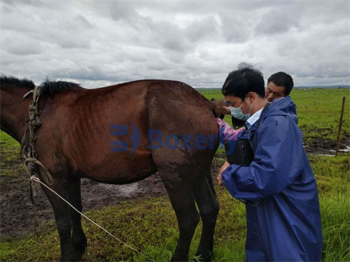
For breeders, veterinarians, and equine farm managers, ensuring the health of a pregnant mare is a task of paramount importance. The welfare of the unborn foal, the longevity of the broodmare’s reproductive career, and the overall economic viability of the breeding program depend on careful monitoring throughout gestation. Among the various diagnostic tools available, ultrasound technology has emerged as the most effective, non-invasive, and insightful method for monitoring the pregnancy status and, critically, the health of the placenta in mares.
In this article, we will explore how ultrasound pregnancy tools are used to assess placental health in pregnant mares, why this monitoring is essential, and how modern breeding farms, particularly in the U.S., Europe, and Australia, have incorporated these techniques into their standard protocols.
Understanding Placental Health in Mares
The placenta plays a vital role in supporting the developing foal, facilitating the exchange of nutrients, gases, and waste products between mare and fetus. Any compromise in placental health—such as infections, inflammation, or abnormalities in size—can lead to serious complications including abortion, premature birth, or compromised neonatal health.
Unlike many other domestic animals, the equine placenta is diffuse, covering nearly the entire surface of the uterus, which makes comprehensive evaluation challenging without specialized tools. Traditional methods such as physical examination or blood markers offer only indirect evidence of placental compromise. Ultrasound technology, however, allows direct visualization of placental structures, providing real-time insights into the uterine environment.
The Evolution of Ultrasound in Equine Reproduction
Ultrasound has been used in equine reproduction since the late 1970s. Initially, its application was largely limited to detecting pregnancy and monitoring early embryonic development. However, as equipment became more sophisticated—offering higher resolution, Doppler capabilities, and portable formats—veterinarians began to use ultrasound extensively for monitoring the placenta throughout gestation.
In countries such as the United States, Australia, and throughout Europe, the incorporation of ultrasound monitoring into equine breeding programs is now standard practice, particularly for high-value sport horses, racing breeds, and endangered genetic lines.
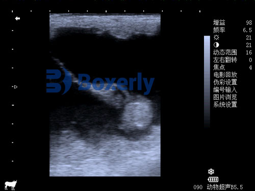
Key Placental Parameters Assessed by Ultrasound
Modern ultrasound pregnancy tools allow veterinarians to evaluate several key aspects of placental health in mares:
1. Combined Thickness of Uterus and Placenta (CTUP):
One of the most widely used measurements, particularly in late gestation, is CTUP. This parameter assesses the combined thickness of the uterine wall and placenta, typically measured at the cervical-placental junction.
-
Normal CTUP values have been well-documented in scientific literature, particularly for breeds like Thoroughbreds and Arabians.
-
An increase in CTUP values beyond breed-specific norms can indicate placentitis (placental infection) or other pathological conditions.
2. Placental Separation or Detachment:
Ultrasound can detect areas where the placenta may be detaching prematurely from the uterine wall, a serious condition that can compromise fetal oxygenation and nutrient transfer.
3. Uteroplacental Blood Flow (via Doppler):
Using Doppler ultrasound, veterinarians can assess blood flow between the uterus and placenta, identifying any vascular insufficiencies that may affect fetal growth.
4. Amniotic Fluid Volume and Quality:
Changes in amniotic fluid—either too much (polyhydramnios) or too little (oligohydramnios)—can reflect placental dysfunction or fetal compromise.
5. Fetal Viability and Growth:
Although not strictly part of the placenta, fetal development can indirectly reflect placental function. Ultrasound allows monitoring of fetal heart rate, activity, and biometric measurements.
Common Placental Problems Detected by Ultrasound
1. Placentitis
Placentitis is among the most common and serious placental diseases in mares, often caused by bacterial or fungal infections ascending through the cervix. Early detection using ultrasound is critical.
-
Ultrasound Signs: Increased CTUP, placental thickening, hyperechoic (bright) lesions, or fluid accumulation.
-
Foreign Perspective: In American and European breeding operations, placentitis is often aggressively monitored with frequent ultrasound checks, particularly in mares with a history of reproductive loss.
2. Premature Placental Separation (Red Bag Delivery)
Ultrasound may detect areas of partial separation long before clinical signs appear at parturition.
3. Hydrops Conditions
Both hydrops allantois and hydrops amnion can be visualized via ultrasound by noting excessive fluid volumes within specific fetal sacs.
4. Twin Pregnancies and Placental Competition
Twin pregnancies in mares pose significant placental challenges. Ultrasound enables early detection and appropriate intervention, such as manual twin reduction, often performed before day 35 of gestation.
When to Perform Ultrasound Placental Monitoring
While every breeding program may develop its own protocol, general guidelines adopted internationally include:
-
Early Pregnancy (10–45 days): Confirm pregnancy, detect twins, assess early embryonic viability.
-
Mid Gestation (5–7 months): Baseline assessment of CTUP and overall uterine environment.
-
Late Pregnancy (7–11 months): Routine monitoring of CTUP, fetal growth, and amniotic fluid levels.
-
High-Risk Pregnancies: Mares with prior abortions, infections, or difficult deliveries may undergo more frequent ultrasound evaluations.
In countries like Australia, where high-performance Thoroughbred racing stock are bred, it’s not uncommon for high-value mares to undergo monthly or even biweekly ultrasound assessments during the final trimester.

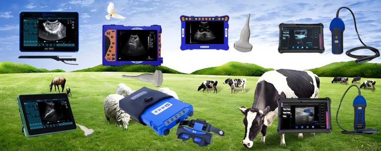
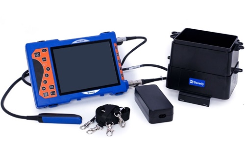
The Role of Advanced Ultrasound Equipment
Modern veterinary ultrasound machines offer several features that are particularly valuable for monitoring placental health:
-
High-Resolution B-Mode Imaging: Critical for detailed assessment of placental thickness and uterine structure.
-
Color Doppler: Allows for real-time evaluation of uteroplacental blood flow.
-
Portability and Durability: Essential for fieldwork on breeding farms where frequent scanning is necessary.
In recent years, devices like the BXL-V50 Veterinary Ultrasound Scanner have gained popularity among equine veterinarians. With its waterproof design, high-definition screen, and portability, it provides accurate imaging even in demanding farm environments. While not always necessary for simple pregnancy checks, such sophisticated devices are invaluable when monitoring complicated or high-risk pregnancies.
Benefits of Ultrasound-Based Placental Monitoring
The incorporation of ultrasound technology into routine prenatal care for mares offers numerous advantages:
1. Early Detection of Problems
Identifying issues before clinical signs emerge allows for timely intervention, whether through medical treatment, supportive care, or adjustments to management practices.
2. Improved Pregnancy Outcomes
Studies consistently show that regular ultrasound monitoring reduces the incidence of late-term pregnancy losses and neonatal complications.
3. Cost-Effective for High-Value Breeding Programs
While some may view frequent ultrasound scans as costly, the potential savings from preventing foal loss or maintaining the reproductive soundness of a valuable mare often outweigh the expense.
4. Non-Invasive and Stress-Free
Unlike more invasive diagnostic methods, ultrasound is well-tolerated by mares and can be performed quickly without sedation or extensive restraint.
5. Real-Time, Quantifiable Data
Veterinarians can document precise measurements and track trends over time, providing an objective basis for clinical decision-making.
Global Adoption and Case Studies
United States:
Large commercial breeding farms in Kentucky and Florida routinely employ ultrasound to monitor placental health, particularly in high-stakes Thoroughbred and Standardbred breeding programs.
Europe:
In Germany, the UK, and France, ultrasound has become indispensable not only for commercial breeders but also for managing endangered native horse breeds where each pregnancy carries genetic significance.
Australia:
The Australian breeding industry, particularly in regions like New South Wales and Victoria, heavily relies on ultrasound tools to protect high-value foals destined for competitive racing markets in Asia and the Middle East.
Case Study – Kentucky Thoroughbred Farm:
On one Kentucky farm, routine CTUP monitoring with ultrasound revealed early signs of placentitis in a valuable mare carrying a high-priced foal. Early antibiotic therapy and strict stall rest led to the successful delivery of a healthy colt, demonstrating the life-saving value of early placental monitoring.
Challenges and Limitations
While ultrasound is immensely valuable, it is not without limitations:
-
Operator Dependency: The accuracy of measurements like CTUP depends heavily on the skill and experience of the ultrasonographer.
-
Interpretation Variability: Some findings may be subtle, and interpreting their clinical significance often requires correlating ultrasound data with laboratory findings and the mare’s history.
-
Equipment Cost: High-end machines with Doppler capabilities can be expensive, which may limit accessibility for small breeders.
Despite these challenges, ongoing advancements in training and equipment are rapidly minimizing these obstacles, making ultrasound monitoring more accessible worldwide.

Future Directions: Artificial Intelligence and Telemedicine
Emerging technologies promise to further enhance ultrasound-based placental monitoring:
-
AI-Based Image Analysis: Algorithms can now assist in automatically measuring CTUP, detecting subtle abnormalities, and standardizing evaluations across operators.
-
Remote Consultation: Cloud-based systems allow veterinarians to share ultrasound images with specialists across the globe, ensuring expert input even in remote or under-resourced locations.
These developments, already being trialed in parts of Europe and North America, suggest that equine prenatal care will continue evolving rapidly, with ultrasound at the center.
Conclusion
Ultrasound pregnancy tools have revolutionized how veterinarians monitor placental health in pregnant mares. With its ability to provide real-time, detailed, and non-invasive assessments, ultrasound empowers breeders to detect problems early, intervene effectively, and safeguard both mare and foal.
As breeding programs become increasingly sophisticated worldwide, especially in high-value racing and sport horse industries, the integration of advanced ultrasound technologies will likely become even more widespread. Tools like the BXL-V50 offer new levels of portability, image clarity, and functionality that align perfectly with the needs of modern equine reproductive medicine.
In the end, protecting placental health is not just about securing a live foal; it is about preserving the genetic future of valuable horses, ensuring welfare standards, and maintaining the economic sustainability of breeding operations. With ultrasound, breeders and veterinarians now have a powerful tool to meet these goals with greater confidence than ever before.
Reference Sources:
-
Bailey, C. S., & McCue, P. M. (2017). “Monitoring the Placenta of Pregnant Mares Using Ultrasonography.” Equine Veterinary Education, 29(5), 275-282.
https://beva.onlinelibrary.wiley.com/doi/full/10.1111/eve.12682 -
Renaudin, C. D., Troedsson, M. H. T., & Pozor, M. A. (2018). Clinical Use of Ultrasonography in Monitoring High-Risk Pregnancies in Mares.” Journal of Equine Veterinary Science, 66, 77-85.
https://www.j-evs.com/article/S0737-0806(18)30245-4/fulltext -
American Association of Equine Practitioners (AAEP). (2023). “Placental Health Monitoring in Mares.”
https://aaep.org