Use of Veterinary Color Doppler Ultrasound
Veterinary diagnostics have made significant strides in recent decades, and one of the most powerful advancements is the integration of Color Doppler ultrasound into routine and emergency practice. This non-invasive imaging technology allows veterinarians to assess blood flow and internal organ function without surgery or causing pain to the animal. Particularly useful in large animal medicine, Color Doppler ultrasound empowers vets to make timely and accurate clinical decisions, even when the patient cannot express symptoms.
This article explores how Color Doppler ultrasound works, its clinical applications in livestock and companion animal medicine, and the practical advantages it offers veterinary professionals and farm operators.
How Color Doppler Ultrasound Works
At its core, Color Doppler ultrasound is based on the Doppler effect—a phenomenon where sound waves change frequency when they bounce off moving objects, such as blood cells. This allows the machine to measure the speed and direction of blood flow in real time.
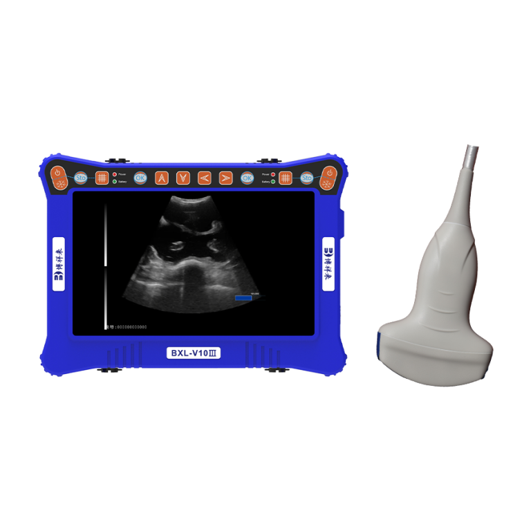
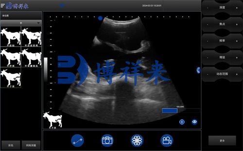
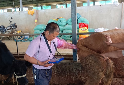
The examination begins with the application of a specialized, conductive gel to the animal’s skin over the target area. This gel eliminates air pockets and enables the ultrasound waves to penetrate the tissue. A transducer—a handheld probe—is then moved gently over the skin, transmitting high-frequency sound waves into the body. These waves reflect off internal structures and are received back by the probe. When the waves encounter moving blood cells, their frequency shifts. This shift is processed by the machine’s computer to create real-time images in color that show the direction and velocity of blood flow.
In animals, where clinical examination is often limited by the inability to communicate symptoms, this visual feedback provides invaluable diagnostic insight.
Applications of Color Doppler in Veterinary Medicine
-
Cardiovascular Diagnostics
Color Doppler ultrasound plays a critical role in evaluating the heart and associated vessels. Veterinarians use this technology to assess cardiac output, detect valve abnormalities, and visualize turbulent or restricted blood flow. This is particularly useful for identifying congenital heart defects or degenerative diseases such as valvular insufficiency in older animals. In large animals like cattle and horses, it helps detect pericardial effusion or cardiac tumors—conditions that would otherwise be nearly impossible to identify in the field without invasive methods.
-
Detection of Vascular Obstructions
One of the hallmark advantages of Color Doppler is its ability to detect vascular obstructions. Blood clots, tumors, or inflamed tissues can impede circulation, resulting in clinical signs such as lameness, swelling, or respiratory difficulty. Color Doppler ultrasound enables early detection of conditions like deep vein thrombosis (DVT) in limbs or pulmonary embolism due to clots traveling to the lungs. Prompt diagnosis allows for faster treatment and improved outcomes.
-
Tumor and Mass Evaluation
Unlike standard grayscale ultrasound, Color Doppler adds functional information to the anatomical structure. It can show whether a mass is vascularized—a key indicator of malignancy. Highly perfused tumors often show abnormal or chaotic blood flow patterns, while benign cysts or fibrous tissue may show minimal or no blood supply. This helps veterinarians prioritize biopsy or surgical removal and determine malignancy risk without needing advanced imaging like CT or MRI.
-
Pregnancy and Reproductive Monitoring
Color Doppler is also invaluable in reproductive medicine. By visualizing blood flow in the uterus and placenta, veterinarians can assess fetal viability, placental integrity, and uterine health. In pregnant mares or cows, Color Doppler can help identify abnormal blood flow that could signal fetal distress or placental insufficiency—conditions that, if not addressed, can result in pregnancy loss.
Furthermore, this technology can help in the diagnosis of ovarian or uterine abnormalities in breeding animals, improving reproductive success rates in farm operations.
-
Liver and Kidney Evaluation
The liver and kidneys are vital organs that can be evaluated using Color Doppler to assess perfusion and detect abnormalities such as tumors, cysts, or infarcts. In cases of suspected hepatic or renal failure, changes in blood flow patterns often precede anatomical changes, allowing earlier intervention.
For example, portal hypertension in the liver can be detected via altered portal vein flow, while renal artery stenosis can signal early kidney dysfunction. These conditions are often silent until advanced stages, making early detection through Doppler ultrasound crucial.
-
Monitoring of Limb Injuries and Inflammation
In animals with lameness or suspected soft tissue injury, Doppler ultrasound can identify increased vascular activity associated with inflammation or infection. This is especially useful in horses, where tendon injuries can be career-limiting. Doppler can guide treatment and monitor healing over time, enabling more precise rehabilitation plans.
-
Guiding Interventions
Color Doppler also assists in performing ultrasound-guided procedures such as fine-needle aspiration (FNA) or biopsy. By identifying major blood vessels in the area, veterinarians can avoid causing hemorrhage, improving the safety and accuracy of diagnostic sampling.
Clinical Advantages for Veterinary Practice
Faster Diagnosis and Immediate Decision-Making
Animals can’t verbalize symptoms, and external signs often appear late in disease progression. With Color Doppler ultrasound, veterinarians can detect abnormalities early and in real time. Whether in a clinic or in the field, this translates into faster treatment and better animal outcomes.
Non-Invasive and Pain-Free
Traditional diagnostic methods like exploratory surgery or radiographic contrast studies are invasive, time-consuming, and often stressful for the animal. Color Doppler is completely non-invasive and typically does not require sedation—especially for smaller animals or those accustomed to handling. In larger livestock, light restraint or mild sedation may be used, depending on the animal’s temperament and condition.
Improved Reproductive Success
In livestock farming, reproductive efficiency directly impacts profitability. Color Doppler allows for detailed examination of reproductive organs and fetal development, helping identify animals with reduced fertility and guiding breeding decisions. It also improves timing for artificial insemination or embryo transfer by monitoring follicular blood flow and uterine perfusion.
Enhanced Client Communication
The color imaging produced by Doppler ultrasound is easy to interpret and can be shared with animal owners to help explain a diagnosis. Clear visuals improve client understanding and trust, leading to higher treatment compliance and better veterinary-client relationships.
Challenges and Considerations
While the benefits of Color Doppler are numerous, its use requires specialized training. Interpretation of Doppler signals, angles, and flow patterns takes time to master. Additionally, the equipment is generally more expensive than conventional grayscale ultrasound systems.
Moreover, environmental factors such as movement, fur thickness, or ambient temperature can affect the quality of imaging. In large animals, full-body scanning may be challenging in field conditions, so preparation and proper restraint are crucial.
Conclusion
Color Doppler ultrasound has emerged as one of the most valuable diagnostic tools in veterinary medicine, offering a dynamic view of internal physiological processes that was once only available through invasive or less precise methods. From cardiac assessment and tumor detection to reproductive monitoring and emergency diagnostics, Color Doppler enhances the veterinarian’s ability to deliver accurate and timely care.
As accessibility and affordability continue to improve, more clinics and mobile veterinary services are expected to adopt this technology. For rural or large-animal practices in particular, it represents a leap forward in animal healthcare and a practical investment in diagnostic excellence.
References
-
DeFrancesco T, Royal K. A survey of point-of-care ultrasound use in veterinary general practice. Education in the Health Professions. 2018;1(2): 50. https://doi.org/10.4103/ehp.ehp_21_18
-
Pelchat J, Chalhoub S, Boyson SR. The use of veterinary point-of-care ultrasound by veterinarians: A nationwide Canadian survey. Canadian Veterinary Journal. December 2020. https://www.ncbi.nlm.nih.gov/pmc/articles/PMC7659883/
-
Lyssens A, Lekane M, Gommeren K, et al. Focused cardiac ultrasound to detect pre-capillary pulmonary hypertension. Frontiers in Veterinary Science. 2022; 9. https://doi.org/10.3389/fvets.2022.830275
-
Feliciano MAR, Uscategui RAR, Maronezi MC, et al. Ultrasonography methods for predicting malignancy in canine mammary tumors. PloS One. 2017; 12(5): e0178143. https://doi.org/10.1371/journal.pone.0178143
-
Pagani E, Tursi M, Lorenzi C, et al. Ultrasonographic features of adrenal gland lesions in dogs can aid in diagnosis. BMC Veterinary Research. 2016; 12(1): 267. https://doi.org/10.1186/s12917-016-0895-1