Veterinary Ultrasound Pregnancy Scanning Evaluating Ovarian Follicular Dynamics
Veterinary practice has long relied on advanced imaging for reproductive management. Among the various diagnostic tools, ultrasound emerges as the most reliable, non‑invasive method for monitoring ovarian follicular dynamics in livestock. From detecting early pregnancy to understanding complex ovarian behaviours, ultrasound scanning offers invaluable insights. This article explores how pregnancy scanning using ultrasound helps evaluate ovarian follicular patterns, drawing from research and practice in Europe, North America, Brazil, India, and beyond.
Follicular Waves and Ultrasonographic Detection
Understanding Follicular Waves
Research has shown that mammals—including cattle, buffaloes, and canines—exhibit follicular wave patterns, where cohorts of follicles grow synchronously, one becomes dominant, then either ovulates or regresses. In bovines, typically two or three such waves occur during each estrous cycle. The ability to detect and monitor these waves is critical for optimizing breeding and insemination timing.
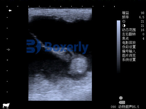
What Ultrasound Reveals
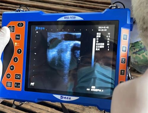
Veterinary-grade ultrasound systems can resolve follicles as small as 2–3 mm in diameter; major antral follicles are easily visualized. Real-time ultrasound enables tracking of follicular growth, regression, and dominance. Sequential scans reveal recruitment, selection, and dominance phases, allowing clinicians to pinpoint ovulation and plan interventions such as timed artificial insemination (TAI).
Pregnancy Scanning and Ovarian Dynamics
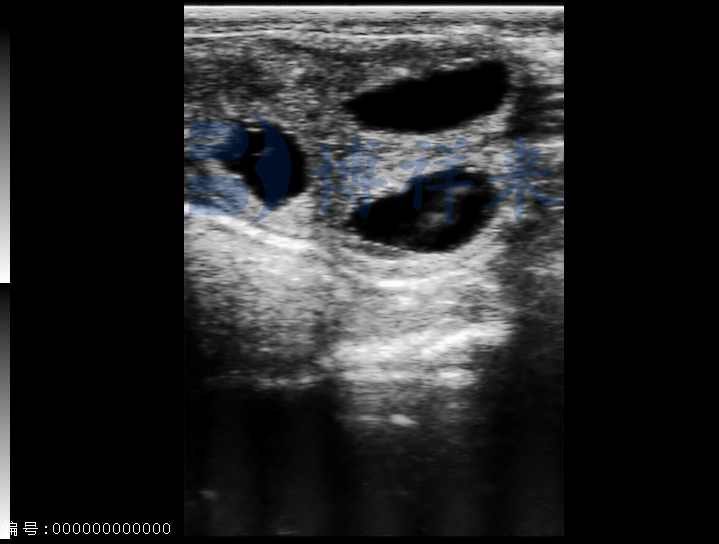
Early Pregnancy Diagnosis
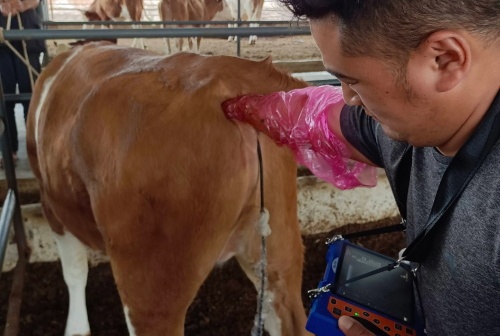
Beyond detecting ovarian structures, ultrasound is a powerful tool for early pregnancy diagnosis. Transrectal scanning can identify fetal vesicles as early as 25–30 days post-breeding in cattle, allowing for early decision-making. This early detection reduces economic loss and improves herd management.
Monitoring Follicular Behavior During Pregnancy
Ultrasonography has revealed that ovarian follicular waves continue during early pregnancy and even the postpartum period. Understanding this continuity is vital for reproductive planning, particularly for early resynchronization programs.
Ovarian Follicle Count and Reproductive Success
High vs Low Antral Follicle Count
A pivotal 2020 study on Bos indicus (e.g. Nelore cattle) compared animals with high versus low antral follicle counts (AFC ≥ 45 vs ≤ 15). Findings showed that lower AFC was associated with larger dominant follicles during TAI and higher pregnancy rates (~58%) compared to high-AFC cows (~45%) . This counterintuitive outcome highlights the importance of ultrasound‑based AFC assessment in managing fertility programs.
Genetic and Environmental Influences
Follicle count and dynamics vary between individuals and are influenced by genetics, breed, nutrition, and management. Studies find repeatable AFCs within individuals across cycles, indicating an intrinsic trait.
Clinical Applications Across Species
In Canines
Ultrasound monitoring of canine ovarian follicles supports reproductive management, especially when using hormone-stimulated cycles. Research shows that ultrasound enables staging of follicular development in bitches, facilitating optimal breeding timing .
In Dairy and Beef Cattle
In dairy and beef cattle, ultrasonography supports postpartum monitoring: detection of ovarian cysts, corpus luteum formation, uterine involution, and return to cyclicity. A Mexican study emphasizes ultrasound’s role in detecting subtle reproductive events and pathologies, improving herd reproductive efficiency .
Advances in Ultrasound Technologies
Doppler and 3D Ultrasound
Color Doppler ultrasound enhances evaluation by mapping ovarian blood flow, offering insight into follicle health and corpus luteum function. Meanwhile, 3D ultrasound systems and AI-driven software enable automated measurement and segmentation, making follicle tracking more efficient and accurate.
AI‑Powered Imaging
Emerging AI methodologies are being applied to ultrasound imaging. Deep learning enables improved beamforming, clutter removal, and super‑resolution imaging. Automated detection and counting of follicles is now possible, enhancing accuracy and reducing user variability .
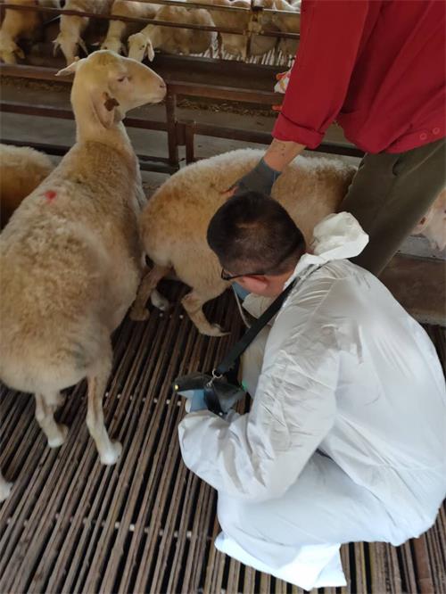
Practical Workflow
-
Initiate baseline scan at estrus or postpartum to count follicles, locate corpus luteum, and assess initial ovarian condition.
-
Perform serial scans (e.g. every 2–3 days) to track follicular wave progression, measure dominant follicle growth, and identify ovulation timing.
-
Scan for pregnancy ~25–35 days post-AI to detect embryo or gestational sac.
-
Rescan throughout gestation to confirm fetal viability and examine ovarian structures like accessory corpora lutea.
Benefits and Challenges
Benefits
-
Non‑invasive: No harm, stress-free for the animal.
-
Accurate tracking: Enables quantitative data on follicle size, number, and growth rate.
-
Informed management decisions: Facilitates timing for AI, embryo transfer, culling or retaining animals.
-
Improved pregnancy outcomes: Early detection allows timely intervention.
Challenges
-
Equipment cost: High-quality veterinary ultrasound devices are expensive.
-
Skill requirement: Operators need training to correctly identify structures and interpret data.
-
Variability: Inter-observer differences can affect accuracy—AI tools and standardization help address this.
Integrating International Perspectives
-
Canada and UK: Adoption of real-time ultrasonography for wave detection and ovulation timing is widespread.
-
Brazil: Large-scale studies on Nelore cattle employing ultrasound-guided TAI and AFC comparisons informed local breeding protocols.
-
India: Genetic and physiological research on follicular wave dynamics underpins breed-specific reproductive strategies .
-
Mexico: Reviews emphasize ultrasound as a core tool in dairy management and reproductive health.
Conclusion
Ultrasound scanning of ovarian follicles and pregnancy status is a cornerstone of modern veterinary reproductive management. By combining real‑time imaging, serial interventions, and emerging AI tools, practitioners can effectively monitor fertility, improve reproductive outcomes, and maximize livestock productivity.
The international body of research underscores the value of ultrasound across breeds and environments. As technologies advance, integrating AI, 3D imaging, and Doppler flow assessment, veterinary professionals will continue to push frontiers in reproductive efficiency.
References
-
Rajamahendran R., Ambrose DJ., Burton B. Clinical and research applications of real‑time ultrasonography in bovine reproduction: a review. Can Vet J. 1994 Sep;35(9):563–72. (PubMed PMID: 7994719) https://www.ncbi.nlm.nih.gov/pubmed/7994719
-
de Lima MA et al. Ovarian follicular dynamics, progesterone concentrations, pregnancy rates and transcriptional patterns in Bos indicus females with a high or low antral follicle count. Sci Rep. 2020;10:19557. https://doi.org/10.1038/s41598-020-76601-5
-
Torres‑Lechuga ME, González‑Maldonado J. Ultrasonography and physiological description of essential events for reproductive management in dairy cattle. Rev Mex Cienc Pecu. 2022;13(2):452–72. https://doi.org/10.22319/rmcp.v13i2.5789
-
DesCôteaux L., Gnemmi G., Colloton J. Ultrasonography of the bovine female genital tract. Vet Clin North Am Food Anim Pract. 2009 Nov;25(3):733–52.
-
Scaled studies on AI and ultrasound: Royo P. et al. Three‑dimensional ultrasound‑based online system for automated ovarian follicle measurement. ArXiv. 2024. https://arxiv.org/abs/2407.18818