Veterinary Ultrasound Tech Monitoring Corpus Luteum Development During Controlled Breeding
In modern livestock reproduction management, precision is paramount. Controlled breeding programs—such as timed artificial insemination (TAI) and synchronization protocols—have revolutionized reproductive efficiency in cattle, sheep, goats, and other food‑producing animals. Central to these programs is the reliable assessment of ovarian structures, particularly the corpus luteum (CL), which governs progesterone production and, in turn, pregnancy establishment and maintenance. Veterinary ultrasound technology, especially transrectal B‑mode ultrasonography, provides a non‑invasive, real‑time window into CL development. This article explores how veterinary ultrasound is applied to monitor CL growth and function during controlled breeding, integrating international best practices and insights.
Understanding the Corpus Luteum and Its Role in Controlled Breeding
Physiology of the Corpus Luteum
After ovulation, the remnant follicular wall transforms into the CL—a transient endocrine gland. Its main role is to secrete progesterone, which prepares the uterine lining for embryo implantation and inhibits further estrous cycles. Inadequate CL development or premature luteolysis can lead to suboptimal progesterone levels, compromising conception rates.
Controlled Breeding Protocols
Protocols such as the Ovsynch (GnRH–PGF₂α–GnRH) in cattle and CIDR‑PGF₂α combinations in small ruminants synchronize ovulation across a herd. Timing of hormone injections hinges on the presence and maturity of the CL:
-
Day 0 (GnRH): Induces ovulation of any dominant follicle and initiates a new follicular wave.
-
Day 7 (PGF₂α): Causes luteolysis of existing CL, dropping progesterone and allowing follicle maturation.
-
Day 9 (GnRH) and Insemination: Triggers ovulation of the synchronized follicle.
Accurate monitoring of CL size and echotexture before and after prostaglandin F₂α (PGF₂α) treatment is critical to confirm luteolysis and predict optimal insemination timing.
Veterinary Ultrasound Technology: Tools and Techniques
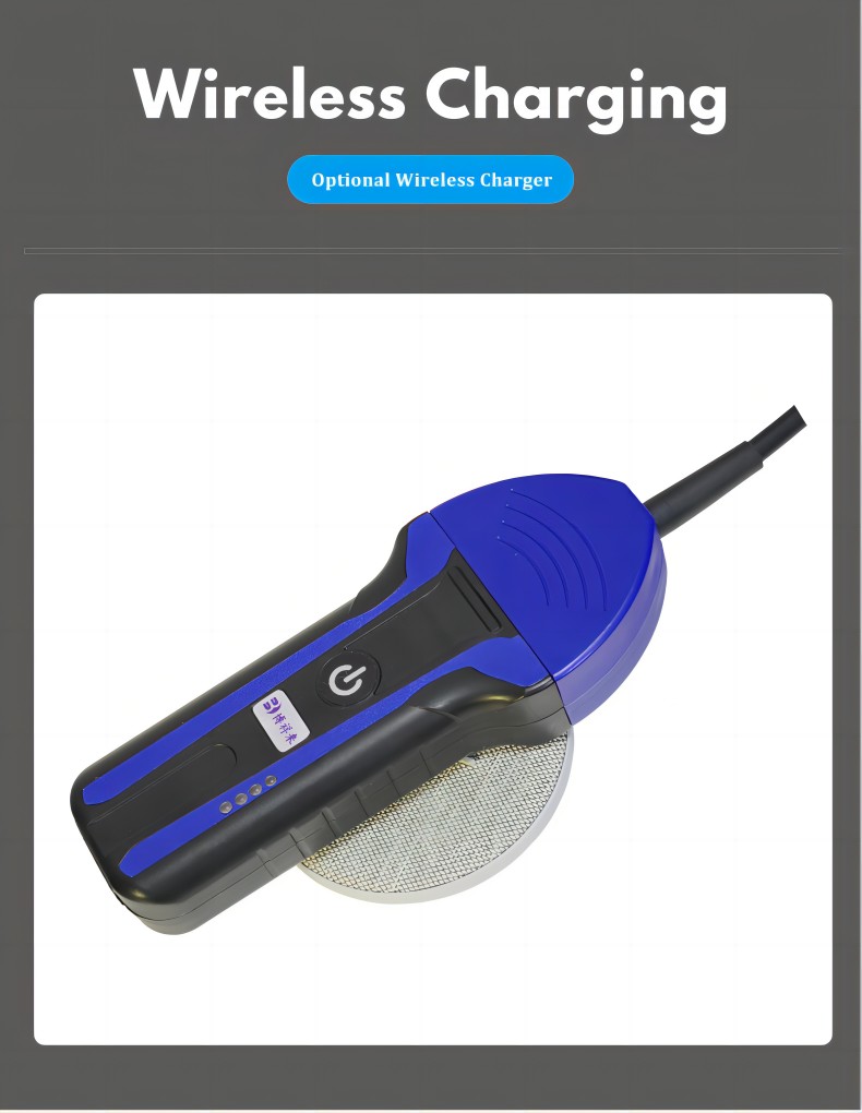
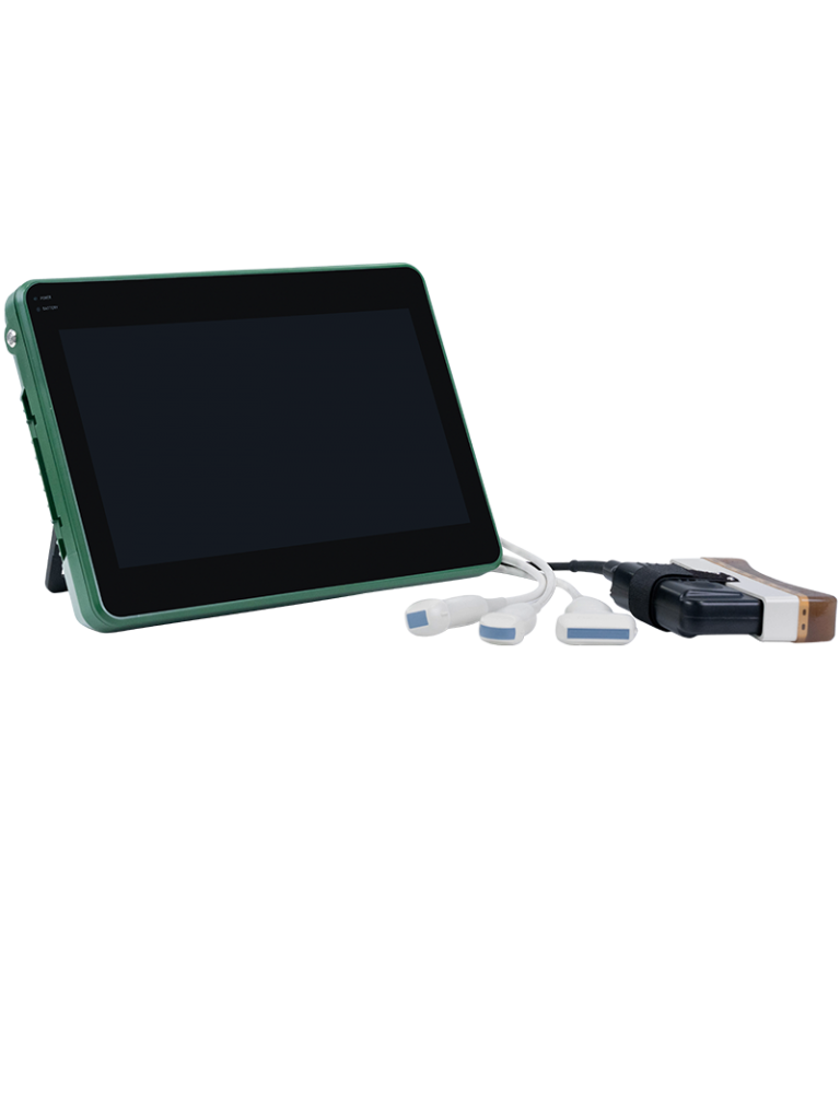
Equipment and Probes
-
B‑mode Ultrasound Scanner: Offers real‑time, gray‑scale cross‑sectional images.
-
Transrectal Linear Probe (5–7.5 MHz): Provides sufficient penetration and resolution for CL measurement in cattle and larger species.
-
Endovaginal Probes (7.5–10 MHz): Used in small ruminants, offering higher resolution for smaller ovarian structures.
Scanning Protocol
-
Preparation: Restrain the animal safely; administer a mild epidural in cattle if needed to relax the rectum.
-
Probe Insertion: Gently introduce the probe transrectally (or transvaginally in sheep/goats), applying coupling gel.
-
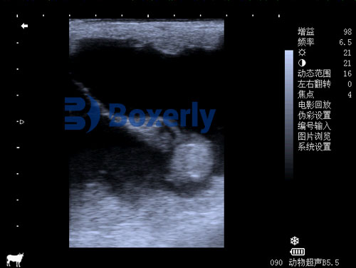
Image Acquisition: Identify the ovary, locate the CL as a rounded, hypoechoic to isoechoic structure on the ovarian surface, and obtain cross‑sectional images.
-
Measurements:
-
CL Diameter: Measure the largest and perpendicular diameters, then calculate mean diameter.
-
CL Area: Trace the outline on frozen images to compute area.
-
CL Volume (optional): Use the ellipsoid formula: (π/6)×length×width×height(π/6) × length × width × height.
-
Monitoring CL Development During Synchronization
Baseline Assessment (Pre‑PGF₂α)
Prior to PGF₂α injection (Day 7), confirm the presence of a functional CL (≥ 17 mm diameter in beef cattle). A well‑developed CL predicts effective luteolysis and synchrony. Studies show that cows with CL diameters < 15 mm at Day 7 have lower conception rates post‑TAI (Silvia et al. 2019) [1].
Luteolysis Confirmation (Post‑PGF₂α)
Approximately 48 hours after PGF₂α administration (Day 9), ultrasound should reveal a significant reduction in CL diameter and echogenicity. A decrease of ≥ 25 % in mean diameter indicates complete luteolysis; persistent CLs warrant rescheduling or additional treatment (Ginther OJ 2018) [2].
Ovulation Verification (Post‑GnRH/Insemination)
On Day 10–11, re‑scan to detect the newly formed CL. Early CL diameter on Day 12 correlates positively with circulating progesterone levels and pregnancy success (Silvia et al. 2019) [1]. Optimal post‑insemination CL size is ≥ 16 mm in dairy cows and ≥ 18 mm in beef breeds.

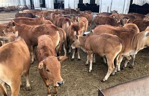
Case Study: Beef Cattle Timed AI Program
At GreenPastures Ranch (U.S. Midwest), a herd of 200 Angus‑cross cows undergoes Ovsynch each 60 days. Ultrasound monitoring workflow:
-
Day 7 Scan: Cows lacking a functional CL (< 17 mm) are removed from TAI and monitored for natural estrus.
-
Day 9 Scan: 180 cows confirm luteolysis; 20 cows (11 %) exhibit persistent CLs and receive a second PGF₂α dose.
-
Day 12 Scan: 165 cows present a new CL ≥ 16 mm and are inseminated; average CL diameter is 18.2 ± 1.3 mm.
-
Pregnancy Check (Day 30): Conception rate is 58 %; non‑pregnant cows’ CL measurements were significantly smaller (mean 14.7 mm), guiding protocol refinements for the next cycle.
Advantages of Ultrasound‑Based CL Monitoring
-
Non‑invasive, Stress‑Free: No surgical procedures; minimal animal handling stress.
-
Real‑Time Decision Making: Immediate data on CL status allows rapid management adjustments.
-
Improved Fertility Outcomes: Identifying sub‑optimal CLs before insemination enhances overall conception rates by 10–15 % compared with hormone protocols alone.
-
Cost‑Effectiveness: Reduces wasted semen and hormone doses on non‑responsive animals.
-
Research Integration: Data supports genetic and nutritional research linking CL characteristics with herd fertility.
Limitations and Considerations
-
Operator Skill: Accurate CL identification and measurement require training; inexperienced operators may misclassify luteal structures.
-
Equipment Costs: High‑resolution scanners represent a significant investment; however, cost per examination decreases with scale.
-
Species‑Specific Protocols: Sheep and goats often require endovaginal scanning and adjusted hormone timelines.
-
Environmental Factors: Rectal temperature and gut contents can affect image quality; consistent scanning conditions are essential.
Future Directions
Advances in portable ultrasound, automated image analysis, and integration with handheld progesterone biosensors promise to further streamline CL monitoring. Machine‑learning algorithms may soon enable on‑device, real‑time luteal classification, reducing reliance on human expertise and expanding adoption in developing regions.
Conclusion
Veterinary ultrasound technology has become indispensable in controlled breeding programs, providing a clear, quantitative view of corpus luteum development. By incorporating routine CL monitoring into synchronization protocols, producers can maximize reproductive efficiency, improve herd fertility, and optimize resource use. As equipment becomes more accessible and analytical tools more sophisticated, ultrasound‑guided breeding management will continue to evolve, benefiting livestock industries worldwide.
References
-
Silvia, W. J., Fricke, P. M., & Bishop, F. M. (2019). Transrectal Ultrasonography of the Corpus Luteum in Cattle: Predicting Fertility Following Timed AI. Journal of Dairy Science, 102(5), 4751–4762. https://doi.org/10.3168/jds.2018-15118
-
Ginther, O. J. (2018). Ultrasonic Imaging and Animal Reproduction (2nd ed.). Wiley. https://www.ncbi.nlm.nih.gov/books/NBK535362/