Ultrasound Pregnancy Tools Detecting Uterine Abnormalities in Breeding Bulls’ Partners
For livestock producers and veterinarians alike, reproductive efficiency is one of the most critical factors influencing farm productivity and profitability. While the fertility of breeding bulls is often closely monitored, it is equally essential to assess the reproductive health of their partners — the females they are bred with. One of the most advanced and reliable methods for evaluating the reproductive health of female breeding animals is the use of ultrasound pregnancy tools. These tools not only confirm pregnancy but also allow for the early detection of uterine abnormalities that can compromise breeding outcomes.

In this article, I’ll explore how ultrasound technology — particularly transrectal and transabdominal ultrasonography — is used to detect uterine abnormalities in breeding bulls’ partners. We’ll review the types of abnormalities that can be identified, how early diagnosis benefits herd management, and why ultrasound has become a cornerstone of reproductive monitoring in modern animal husbandry.
Understanding Reproductive Challenges in Breeding Programs
In any breeding operation, whether involving cattle, sheep, goats, or horses, successful conception depends not just on the male’s fertility but also on the uterine environment of the female. Subclinical uterine abnormalities often go unnoticed through routine visual examinations or behavioral observation. These hidden issues can cause failed conception, early embryonic death, or reduced litter size — all of which reduce farm profitability.
In cattle breeding, for example, studies estimate that up to 20-30% of reproductive failures may be associated with undiagnosed uterine abnormalities. These may include endometritis, uterine cysts, fluid accumulation, fibroids, or adhesions — conditions that can persist even in animals showing outward signs of estrus.
This is where ultrasound technology provides a critical advantage.
The Role of Ultrasound Pregnancy Tools in Breeding Programs
Ultrasound pregnancy tools, particularly portable veterinary units equipped with linear or convex probes, allow real-time imaging of the reproductive tract. In breeding bulls’ partners, these devices are primarily used to:
-
Confirm pregnancy status
-
Assess fetal viability
-
Evaluate the uterine and ovarian structures
-
Detect abnormalities that may hinder fertility or pregnancy maintenance
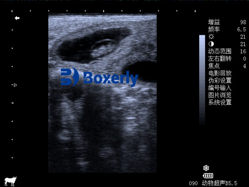
Modern ultrasound equipment, such as transrectal scanners, provides high-resolution, non-invasive visualization of the uterus, ovaries, and developing embryo within weeks of conception. Early detection of uterine abnormalities means timely intervention, ultimately improving reproductive efficiency and economic returns.

Common Uterine Abnormalities Detected by Ultrasound
1. Uterine Fluid Accumulation
The presence of fluid within the uterine cavity can indicate inflammation (endometritis), infection (pyometra), or hormonal imbalances. Using ultrasound, veterinarians can observe hypoechoic (dark) or anechoic (black) areas within the uterus that suggest fluid accumulation. The volume, consistency, and distribution of the fluid help differentiate between pathological conditions.
2. Endometrial Fibrosis and Fibroids
Older females or those with repeated pregnancies may develop fibrous tissue or benign tumors (fibroids) within the uterine wall. These are easily visualized as hyperechoic (bright) masses on ultrasound, sometimes with shadowing artifacts, which may impede normal implantation or fetal development.
3. Uterine Adhesions
Adhesions between uterine horns, ovaries, or the abdominal wall can restrict normal uterine function. These adhesions may follow trauma, surgery, or chronic inflammation and are challenging to diagnose without imaging. Ultrasonography helps identify abnormal tissue connections that compromise uterine mobility and elasticity.
4. Uterine Cysts
While ovarian cysts are more commonly discussed, uterine cysts also occur. They can appear as small, round, fluid-filled structures along the uterine lining and may interfere with embryo implantation if numerous or strategically located.
5. Early Embryonic Loss Indicators
Through repeated ultrasound examinations, veterinarians can monitor embryo development. A shrinking or absent embryo sac, irregular fetal heartbeat, or abnormal placental development may signal early embryonic death — allowing for prompt reproductive management adjustments.
Advantages of Early Ultrasound Diagnosis
The benefits of using ultrasound pregnancy tools extend beyond confirming pregnancy:
-
Timely Interventions: Early diagnosis of uterine abnormalities allows for immediate treatment — whether with antibiotics, hormonal therapy, or surgical correction.
-
Reduced Breeding Delays: Identifying non-pregnant animals or those unsuitable for breeding helps optimize breeding schedules and reduces wasted semen or bull service.
-
Improved Genetic Selection: Females with chronic or untreatable uterine conditions can be culled from the breeding program, ensuring that only healthy, reproductively sound animals contribute to the herd’s genetic base.
-
Enhanced Animal Welfare: Non-invasive, painless ultrasound scans reduce animal stress compared to invasive diagnostic methods.
Real-Life Application: Case Example
On one North American beef cattle farm, veterinarians employed transrectal ultrasound with a linear 7.5 MHz probe during routine pregnancy checks. One valuable cow repeatedly failed to conceive despite multiple mating attempts with a proven bull. Ultrasound revealed fluid pockets in both uterine horns, indicative of subclinical endometritis.
Prompt antibiotic treatment resolved the infection, and within two estrous cycles, the cow conceived successfully. Without ultrasound, this condition might have remained undiagnosed, leading to prolonged infertility, economic loss, and unnecessary culling.
Technological Evolution: Portable Ultrasound Scanners
The development of portable, high-resolution ultrasound devices has revolutionized on-farm reproductive monitoring. Compact systems with waterproof casings, extended battery life, and real-time image storage allow veterinarians and farm managers to conduct thorough evaluations even in challenging field conditions.
While transrectal scanning remains the gold standard for large animals like cattle, smaller species such as goats and sheep often benefit from transabdominal ultrasound, which provides clear imaging even in early gestation.
Veterinary ultrasound manufacturers, including companies developing advanced models like the BXL-V50, offer devices tailored to farm animal reproduction. Such tools combine durability with high imaging precision, allowing accurate detection of both pregnancy and reproductive pathologies in various livestock species.

Ultrasound Versus Traditional Methods
Traditional methods for evaluating reproductive health, such as rectal palpation or hormonal assays, have significant limitations:
-
Palpation: Highly dependent on operator skill; less accurate in early gestation or detecting subtle uterine abnormalities.
-
Blood Tests: Indicate hormonal status but do not reveal physical abnormalities.
-
Visual Observation: Detects only external signs of estrus; many uterine issues remain unnoticed.
In contrast, ultrasound provides a real-time, visual representation of reproductive structures, allowing veterinarians to observe anatomy directly rather than infer it from indirect signs.
The Global Perspective: Why International Farmers Adopt Ultrasound
Across Europe, North America, and Australia, the adoption of ultrasound pregnancy tools in livestock breeding continues to grow. This trend reflects increasing awareness of several global farming realities:
-
Economic Pressures: With high input costs and narrow profit margins, producers require precision tools to minimize reproductive losses.
-
Animal Welfare Standards: Many countries mandate or encourage non-invasive diagnostic approaches to reduce animal stress.
-
Technological Accessibility: Portable ultrasound devices have become more affordable and user-friendly, even for small-scale farmers.
-
Data-Driven Farming: Integrating ultrasound findings into herd management software supports data-based decision-making.
According to a 2023 report from the International Livestock Research Institute, farms utilizing ultrasound for reproductive monitoring reported a 12-18% improvement in overall conception rates compared to those relying solely on conventional methods.
The Veterinary Perspective: Ultrasound in Preventive Reproductive Health
For veterinarians specializing in reproductive management, ultrasound serves as both a diagnostic and preventive tool:
-
Baseline Assessment: Evaluating the uterus before the breeding season identifies animals requiring preemptive treatment.
-
Post-Breeding Checks: Early confirmation of successful fertilization allows rapid resynchronization for non-pregnant animals.
-
Monitoring Treatments: Following medical or surgical intervention, ultrasound helps track recovery progress, ensuring issues do not recur.
In veterinary reproductive practice, ultrasound has transitioned from being a luxury tool to an essential service standard.
Conclusion
In modern livestock breeding operations, achieving high conception rates requires more than just selecting fertile bulls. The reproductive health of breeding females plays an equally crucial role, and undiagnosed uterine abnormalities can quietly erode herd productivity. Ultrasound pregnancy tools offer livestock managers a reliable, non-invasive, and highly informative window into the reproductive status of breeding bulls’ partners.
By detecting uterine abnormalities early, ultrasound enables timely interventions that improve breeding success, enhance animal welfare, and optimize economic returns. As portable ultrasound technology continues to advance, it will undoubtedly become even more integrated into routine reproductive management worldwide — helping farmers and veterinarians work together for healthier, more productive herds.
Reference Sources
-
Sheldon, I.M., Lewis, G.S., LeBlanc, S., & Gilbert, R.O. (2006). Defining postpartum uterine disease in cattle. Theriogenology, 65(8), 1516-1530. https://doi.org/10.1016/j.theriogenology.2005.08.021
-
International Livestock Research Institute (2023). Global Trends in Reproductive Ultrasound Adoption. https://www.ilri.org/reproductive-ultrasound
-
Whitaker, D.A., & Smith, E. (2021). Veterinary Ultrasonography in Food-Producing Animals. Journal of Veterinary Imaging.
-
Beef Cattle Institute (2023). “Use of Ultrasound for Growth Evaluation in Cattle.” https://www.beefcattleinstitute.org/ultrasound-growth