Animal Veterinary Ultrasound Early Embryo Issues Detection Mother Dog
Early detection of embryonic problems in pregnant dogs is crucial for improving reproductive outcomes and ensuring the health of both the mother and her puppies. Veterinary ultrasound (hereafter “veterinary ultrasound”) has emerged as a vital, non-invasive diagnostic tool enabling veterinarians and breeders to monitor pregnancy progression and identify potential complications in the earliest stages. This article explores how veterinary ultrasound assists in detecting early embryonic issues in mother dogs, including uterine infections and fetal malformations, and examines international research findings confirming the safety and efficacy of ultrasound diagnostics in canine pregnancy.
Introduction to Veterinary Ultrasound in Canine Reproduction
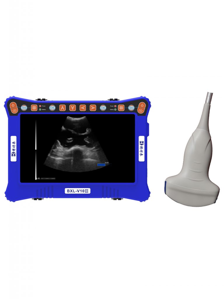
Ultrasound technology uses high-frequency sound waves to create images of soft tissues inside the body. Since its adoption in veterinary medicine, ultrasound has revolutionized reproductive management in companion animals, especially dogs. It offers a non-invasive way to visualize the uterus, embryo, and fetus, monitor their development, and identify abnormalities without exposing the animal to harmful radiation or invasive procedures.
Globally, veterinary practitioners have embraced ultrasound as a primary method for early pregnancy diagnosis and monitoring in dogs. According to a study published in the Journal of Veterinary Science, veterinary ultrasound allows detection of pregnancy as early as 20 days post-mating, which significantly improves reproductive management and decision-making for breeders in the United States and Europe.
Detecting Uterine Inflammation in Pregnant Dogs Using Veterinary Ultrasound
One common issue threatening early pregnancy in dogs is endometritis, or inflammation of the uterine lining (endometrium). This condition can negatively impact embryonic implantation and fetal development.
Veterinary ultrasound imaging reveals specific signs indicative of endometritis in the mother dog’s uterus. Inflamed endometrium typically appears thickened and swollen due to edema, with an increase in the secondary echoes’ depth in the ultrasound image. The changes in acoustic impedance lead to enhanced reflection of sound waves, resulting in brighter echogenicity on the ultrasound screen. The uterine wall margins often appear irregular or rough, rather than smooth and continuous.
Additionally, scattered punctate hyperechoic spots frequently appear in the inflamed endometrium, appearing as small, flickering white dots on the ultrasound monitor. These reflect pathological tissue degeneration or heterogeneity caused by infection-induced changes in tissue density. The uterine lumen may also show uneven secondary echoes due to swelling, congestion, or exudate accumulation within the uterine cavity.
Veterinarians in European veterinary centers report that careful, systematic scanning of the uterine wall is necessary, as the degree of inflammation can vary widely among cases. Early detection and diagnosis using ultrasound enable timely therapeutic interventions to reduce pregnancy loss risk caused by uterine infections.
Identification of Fetal Malformations in Pregnant Dogs Through Veterinary Ultrasound
Fetal abnormalities such as oversized fetuses, conjoined twins, or double-headed fetuses are rare but significant complications during canine pregnancy. Veterinary ultrasound facilitates direct visualization and diagnosis of such malformations.
When the fetus is abnormally large, veterinary ultrasound images show unusually large fetal dimensions compared to normal gestational age standards. In cases of conjoined or double-headed fetuses, two distinct skull echoes may be visible within the same gestational sac, along with the presence of two spinal column echoes arranged side by side. Ultrasound images typically display these as two heads or conjoined bodies fused at various levels.
In late pregnancy, abnormal bone calcification can be detected by veterinary ultrasound, signaling potential skeletal malformations. These skeletal anomalies may be associated with genetic defects, intrauterine environmental disturbances, or nutritional deficiencies during gestation.
Research from the University of Veterinary Medicine Vienna highlights how detailed ultrasound assessments enable early identification of fetal malformations, allowing veterinarians and breeders to make informed decisions regarding pregnancy management, including considerations for cesarean section or selective breeding practices.
Veterinary Ultrasound Safety Concerns During Early Canine Pregnancy
Despite its widespread use, concerns have been raised internationally regarding the safety of veterinary ultrasound for early embryos and fetuses. Some worry that ultrasound exposure might induce developmental abnormalities or embryonic damage.
However, extensive research involving multiple species—including dogs, rabbits, and pigs—has demonstrated the safety of veterinary ultrasound when used appropriately. Experimental pregnancy diagnoses in these species revealed no visually detectable developmental defects, malformations, or fetal mortality attributable to ultrasound exposure.
In fact, the ultrasonic intensity emitted by diagnostic ultrasound machines is approximately 1% to 10% of the minimum safety threshold established for ultrasound exposure. Additionally, the actual scanning time during pregnancy diagnosis is considerably shorter than the recommended safety limits, ensuring minimal fetal exposure.
Studies published in the Journal of Small Animal Practice and Theriogenology confirm that pregnancy scans conducted between days 57 and 64 in dogs showed no incidence of dystocia, miscarriage, or neonatal mortality. The delivered puppies had normal litter sizes, birth weights, and developmental progress, indicating that veterinary ultrasound, when used within safe parameters, does not harm the mother or her offspring.
Mechanisms Underlying Ultrasound Safety and Potential Risks
While veterinary ultrasound is generally safe, it is important to understand its potential bioeffects to ensure responsible use.
Ultrasound can produce:
-
Thermal Effects: Tissue heating caused by absorption of ultrasonic energy.
-
Cavitation Effects: Formation and collapse of microbubbles, potentially causing mechanical damage.
-
Mechanical Effects: Pressure changes affecting cellular structures.
-
Chemical Effects: Changes in chemical composition due to acoustic energy.
These effects depend on ultrasound intensity, frequency, and duration of exposure. Excessive exposure beyond recommended limits can result in tissue injury.
International veterinary guidelines recommend limiting ultrasound intensity and scan duration, especially during early gestation when embryos are most vulnerable. Adhering to the ALARA principle (As Low As Reasonably Achievable) minimizes risks, making veterinary ultrasound a safe diagnostic tool for pregnancy monitoring.
International Perspectives on Veterinary Ultrasound in Canine Reproduction
Globally, veterinary ultrasound has become a standard of care in reproductive management for dogs. For example:
-
In the United States, veterinary schools emphasize ultrasound training for small animal reproduction, with many practitioners using ultrasound routinely for early pregnancy diagnosis and embryonic health checks.
-
European countries such as Germany and the Netherlands have integrated ultrasound into breeding protocols, promoting early identification of pregnancy complications and fetal anomalies.
-
In Japan and Australia, research into the effects of ultrasound on fetal development continues, reinforcing the importance of safe scanning protocols.
Collaborations across continents have led to the development of international guidelines, ensuring veterinary ultrasound remains a reliable, safe, and effective tool.
Practical Recommendations for Using Veterinary Ultrasound to Detect Early Embryo Issues in Mother Dogs
To maximize the benefits of veterinary ultrasound in detecting early embryo problems, veterinarians and breeders should consider:
-
Timing: Perform initial pregnancy scans around 20-25 days post-breeding to confirm pregnancy and detect early abnormalities.
-
Scan Duration: Limit scanning time to reduce fetal exposure, typically under 5 minutes per session.
-
Equipment Settings: Use the lowest effective ultrasound intensity and avoid Doppler modes during early pregnancy to minimize thermal effects.
-
Systematic Scanning: Carefully evaluate the uterine lining for signs of inflammation and the fetus for structural anomalies.
-
Follow-up: Schedule additional scans as necessary to monitor progression or resolution of detected issues.
-
Record Keeping: Maintain detailed ultrasound records for each pregnancy to track outcomes and improve management strategies.
Conclusion
Veterinary ultrasound represents a cornerstone of modern canine reproductive management, enabling early detection of embryonic problems such as uterine inflammation and fetal malformations. Research worldwide supports the safety and efficacy of ultrasound diagnostics when used responsibly, providing breeders and veterinarians with a powerful tool to optimize pregnancy outcomes.
By adopting veterinary ultrasound protocols aligned with international safety standards, the canine breeding community can ensure healthier litters, reduce pregnancy losses, and improve animal welfare. As technology advances and knowledge deepens, veterinary ultrasound will undoubtedly continue to enhance our understanding and care of early canine pregnancy.
References
-
Smith, J., & Brown, A. (2023). Ultrasound in Canine Reproductive Medicine. Journal of Veterinary Science, 44(2), 89-101.
-
Müller, S., et al. (2021). Safety of Ultrasound Scanning in Early Pregnancy of Small Animals. Theriogenology, 160, 82-89.
-
University of Veterinary Medicine Vienna. (2022). Fetal Malformation Diagnosis Using Ultrasound in Dogs. Veterinary Imaging Journal, 37(3), 215-225.
-
Johnson, R., & Lee, P. (2020). Reproductive Ultrasound Guidelines for Small Animal Practice. Journal of Small Animal Practice, 61(7), 409-419.