Methods for Restraining Ewes During Ultrasound Examinations
If you’re planning to ultrasound a ewe, the first thing you need is a calm animal that stays still during the procedure. The way you restrain the ewe can significantly impact both the efficiency and accuracy of the scan. Choosing the right positioning makes the process smoother for both the handler and the animal. Common restraint methods include natural standing, lateral recumbency (lying on the side), or using a dedicated holding frame. The best choice depends on the ewe’s temperament and the purpose of the exam.

Choosing the Right Restraint Position for Easy Handling
The most straightforward method is to scan the ewe while she’s standing. An assistant can gently hold her neck or lightly secure her body with their legs to keep her still. This technique is especially useful in large flocks where moving each animal individually would be too time-consuming. If you have a more restless ewe, a simple restraining frame can help stabilize her and make the process safer and more efficient.
While scanning a ewe in lateral recumbency can offer a slightly earlier pregnancy diagnosis with improved image clarity, it also takes more time and effort to get the ewe down and properly positioned. This method can be useful during early pregnancy checks when certain structures are more visible from the side.
However, with large numbers of animals, the time required makes this method less practical. In real-world practice, whether the ewe is lying on her side, on her back, or standing isn’t a critical factor in early scans. The key is to go with whatever position keeps the ewe calm and comfortable.

Key Details to Watch During the Exam
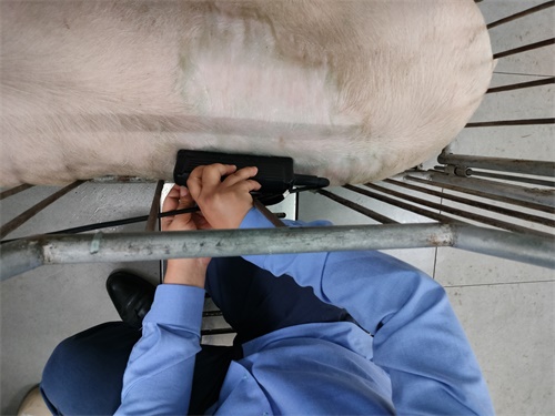
Before you begin, give the ewe a few minutes to settle into the environment. Avoid rushing into the procedure. The handler can gently stroke her neck or pat her back to help her relax. If the ewe is visibly nervous or trembling, the ultrasound image may turn out blurry. In that case, pause and calm her before continuing. When placing the probe on the abdominal area, always apply a layer of clear ultrasound gel to the skin first. This ensures better contact and clearer imaging. Don’t skimp on gel, but there’s no need to overdo it—a thin, even layer works best.

The primary scanning zones are the area on either side of the udder and the lower right abdomen. For early pregnancy diagnosis, spend extra time scanning the less hairy regions around the udder, and move the probe slowly to carefully locate the target structures. If the ewe has a large belly or thick fleece that’s blocking visibility, an assistant can gently lift a hind leg for a better angle. There’s usually no need to shear—adjusting the position often does the trick.

During the scan, make sure the probe stays in constant contact with the skin—don’t let it hover. Move it in a gentle fanning motion rather than in straight lines for better coverage and image quality.
Experienced handlers often reach from behind the ewe to scan, which avoids the need to reposition the animal and allows for quicker targeting. For more excitable ewes, securing the neck with your legs can be less tiring than using your hands, and it frees you up to adjust ultrasound settings as needed. Ideally, the entire scan should be completed in three to five minutes to prevent restlessness, which can lead to poor image quality and inaccurate results.