
Evaluating Reproductive Capacity in Gilts with Ultrasonography
Ultrasound imaging has become an indispensable tool in modern livestock management, especially in swine reproduction. As a farmer focused on pig breeding, I’ve seen how ultrasonography not only provides insight into the health status of animals but also plays a crucial role in assessing reproductive readiness in gilts. This article explores how ultrasound helps evaluate the reproductive capabilities of young female pigs (gilts), the anatomical development behind fertility, and the influence of boar exposure on sexual maturity.

Understanding Reproductive Development in Gilts
From a physiological standpoint, reproductive capacity in gilts is deeply tied to the structural and developmental maturity of their reproductive organs. These structures begin forming early—around 30 to 50 days after fertilization. During this embryonic phase, the ovaries, uterus, and central reproductive control centers in the brain begin to differentiate. Even though they continue developing throughout gestation, an ultrasound scan at this stage reveals relatively immature genitalia. The uterus appears quite small, and the ovaries are not yet active.
Interestingly, by birth, the ovaries already contain all the ova that the gilt will ever release throughout her life. This early formation is critical, as it sets the foundation for future fertility. By the time gilts reach 60 to 90 days of age, cellular specialization within the reproductive tract becomes more defined. Ovarian follicles begin their maturation process, and uterine tissues start developing into functional structures that can support embryo implantation and development. Muscle cells in the uterus also begin forming, which are vital for the farrowing process.
The Role of Ultrasonography in Assessing Reproductive Structures
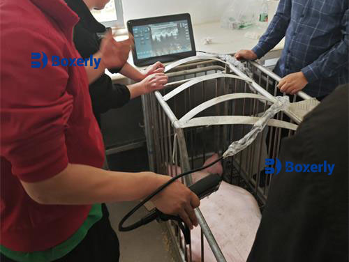
Ultrasound technology enables us to visualize these internal structures in a non-invasive, real-time manner. B-mode (brightness mode) imaging, the most commonly used format, allows us to see grayscale images of the uterus and ovaries. By placing a transducer on the animal and using coupling gel, we can direct ultrasonic pulses into the body. When these pulses hit tissues of different densities, they bounce back, forming images that help us evaluate the size, shape, and internal patterns of organs.
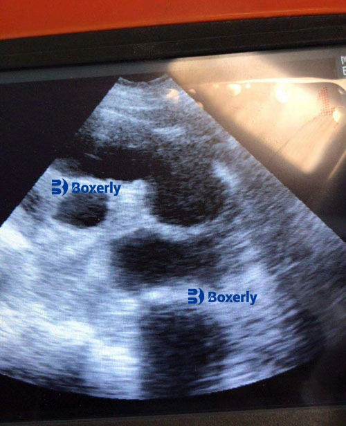
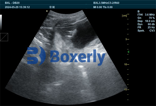
In gilts, ultrasonography provides a way to confirm whether the reproductive system is developing appropriately. We can assess the size of the uterus, the presence and condition of follicles in the ovaries, and even detect early signs of hormonal changes. Gilts with underdeveloped or inactive reproductive organs often have poor breeding outcomes. Thus, early detection helps us decide which animals are best suited for reproduction and which may require additional monitoring.
Influence of Boar Exposure on Reproductive Maturity
One of the most practical and effective strategies for promoting reproductive development in gilts is exposure to mature boars. Starting around 150 days of age, introducing gilts to adult boars (10 months or older) can significantly reduce the age at which gilts reach puberty. This is not only evident behaviorally—through visible signs of estrus and standing reflex—but also physiologically, as seen through ultrasound imaging.
Boar exposure stimulates the gilt’s endocrine system. Boar saliva contains pheromones that trigger hormonal responses in young females, leading to the maturation of ovarian follicles and the thickening of uterine walls. When we scan these gilts with ultrasound, we often observe larger, more mature follicles and an increase in uterine volume compared to gilts raised without boar contact.
In my experience, gilts that respond to the first boar exposure within 28 days—especially with a standing reflex—are typically those with a well-developed reproductive system. These animals also tend to have a longer reproductive lifespan. Thus, the presence or absence of this early response can help us predict future breeding performance.
Correlation Between Growth Rate and Reproductive Potential
The connection between body growth and reproductive ability is another area where ultrasound has proven valuable. Gilts that grow rapidly and reach a healthy body condition between 160 and 220 days of age are more likely to respond to boar exposure and show signs of estrus. Ultrasound can validate this by showing progressive development in ovarian follicles and uterine structures.
Selecting replacement gilts based on both visual inspection and ultrasonographic evidence ensures we retain only the most fertile individuals in the breeding herd. By correlating body condition scores, behavioral signs of estrus, and internal reproductive metrics, we improve our chances of successful mating and reduce non-productive days.
Benefits of Early Ultrasonographic Evaluation
Using ultrasound in early gilt selection offers numerous advantages:
-
Precision Breeding: We can more accurately select gilts ready for breeding, improving farrowing rates.
-
Reduced Costs: By avoiding the breeding of underdeveloped or infertile gilts, we save time and resources.
-
Longer Reproductive Lifespan: Identifying high-potential gilts early helps us maintain a productive and efficient breeding herd.
-
Increased Predictability: When combined with boar exposure protocols, ultrasound provides clear evidence of puberty onset and ovarian activity.
Practical Tips for Implementation
Here are a few tips I follow on my farm when using ultrasound for gilt reproductive evaluation:
-
Start early: Begin evaluating gilts around 150 days of age.
-
Use consistent boar exposure: Daily exposure to adult boars increases the reliability of ultrasound findings.
-
Keep detailed records: Document growth rates, estrus signs, and ultrasound results for each gilt.
-
Monitor follicular development: Pay attention to the size and number of follicles on each ovary.
-
Reassess regularly: Scan animals more than once if necessary to confirm findings.
Conclusion
Ultrasonography is more than a diagnostic tool—it’s a management strategy that helps improve reproductive outcomes in pig farming. By integrating ultrasound into our daily routines, we gain a clearer understanding of gilt development and fertility. Coupled with boar exposure, proper nutrition, and environmental management, ultrasound ensures that every gilt selected for breeding has the highest chance of success. For those of us dedicated to efficient and sustainable pig production, it’s a technology we simply can’t afford to ignore.


Read this during lunch—super interesting stuff. Never thought ultrasounds were used like this in pigs. The part about tracking follicle size really caught my eye. Makes way more sense now why early detection matters. Cool read.
Gostaria de entender melhor como funciona essa avaliação por ultrassom nas porcas jovens. Alguém já usou esse tipo de tecnologia? Dá pra aplicar em propriedades menores? Estou pensando em investir, mas queria ouvir experiências reais primeiro.
This is exactly the kind of info we need on the farm. I’ve been using ultrasound on gilts for a while, but never thought much about how early follicle development could guide breeding decisions. The part about differentiating fluid patterns in the uterus was especially helpful.