Veterinary Ultrasound Tech Evaluating Uterine Pathology Risks In Postpartum Sows
As a swine producer, ensuring the rapid and complete recovery of sows after farrowing is paramount for maintaining herd productivity and minimizing economic losses. Among the array of diagnostic tools available, ultrasonography—specifically B‑mode ultrasound—has emerged as a non‑invasive, real‑time, and highly informative method for evaluating uterine involution, detecting early pathological changes, and assessing risks such as endometritis or retained placental fragments in postpartum sows. In this article, I will share how veterinary ultrasound technology is applied on commercial pig farms to monitor uterine health, discuss key sonographic parameters, and explain why this approach has gained traction among swine specialists worldwide.

Role of Postpartum Uterine Monitoring in Sow Productivity
Importance of Early Detection
Postpartum uterine pathologies—most notably metritis and endometritis—can significantly impair subsequent reproductive performance, leading to delayed return to estrus, reduced conception rates, and lower litter sizes. Early identification of such conditions allows for timely intervention, whether antimicrobial therapy or drainage procedures, thereby reducing culling rates and preserving lifetime productivity of valuable breeding sows.
Physiological Uterine Involution
Following farrowing, the sow’s uterus undergoes a process of involution during which endometrial repair, uterine size reduction, and lochia clearance occur. Under optimal conditions, complete involution may take three to four weeks. However, environmental stressors, farrowing complications, or inadequate postnatal care can delay involution and predispose to bacterial proliferation.
Ultrasonographic Assessment of Postpartum Uterine Health
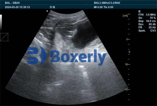
B‑Mode Imaging Technique
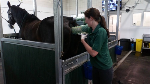
Using a high‑frequency linear transducer (5–7.5 MHz), ultrasound tech can be performed transabdominally on sows restrained in a farrowing crate. The transducer is placed caudal to the last pair of ribs, scanning in both sagittal and transverse planes to visualize the uterine horns, body, and endometrial lining.
Key Sonographic Indicators
-
Uterine Diameter and Wall Thickness
-
Normal involuting horn diameter decreases from approximately 4–5 cm on day 1 postpartum to < 2 cm by day 21.
-
Increased wall thickness (> 8 mm) beyond day 7 may indicate inflammation or edema.
-
-
Intrauterine Fluid Accumulation
-
Minimal anechoic fluid (< 1 cm) is physiological in early involution.
-
Persistent or echogenic fluid pockets suggest purulent exudate or retained placental tissue.
-
-
Endometrial Irregularities
-
Hyperechoic debris adherent to the endometrium often corresponds to necrotic tissue or fibrin.
-
Focal masses may represent retained fragments requiring manual removal.
-
-
Doppler Evaluation (when available)
-
Color‑Doppler can assess uterine blood flow; reduced perfusion may reflect ischemia, whereas hyperemia often accompanies active inflammation.
-
Protocol for Routine Postpartum Scanning
-
Timing
-
Day 3–5: Establish baseline uterine size and fluid status.
-
Day 10–14: Monitor involution progress; detect delayed clearance.
-
Day 21–28: Confirm return to near‑baseline dimensions before breeding.
-
-
Animal Handling
-
Minimal restraint; gentle cleaning of flank with alcohol.
-
Use of ultrasound gel ensures optimal acoustic coupling.
-
-
Data Recording
-
Document uterine horn diameters, fluid measurements, and wall thickness in herd records.
-
Correlate findings with rectal temperature, feed intake, and farrowing complications.
-
Case Studies and Field Experiences
Case 1: Early Metritis Detection
On a 500‑sow commercial unit in Spain, routine scans on day 7 postpartum identified eight sows with uterine wall thicknesses > 9 mm and echogenic fluid pockets. Early administration of systemic antibiotics and intrauterine lavage resulted in 100 percent recovery without prolonged wean‑to‑service intervals.
Case 2: Retained Placenta Complication
A high‑performance multiplier herd in Denmark reported sporadic retained placentas at farrowing. Ultrasound at day 14 postpartum revealed persistent hyperechoic masses in four sows. Manual removal under sedation coupled with targeted antimicrobial therapy restored normal involution by day 21, compared to historical average of 35 days.
Advantages of Ultrasound Surveillance
-
Non‑Invasive and Stress‑Free: Unlike manual palpation or exploratory surgery, ultrasonography poses no additional stress and can be repeated frequently.
-
Quantitative and Objective: Provides measurable data on uterine dimensions and fluid volumes, reducing reliance on subjective clinical signs.
-
Real‑Time Decision Making: Immediate visualization facilitates on‑the‑spot intervention planning.
-
Enhanced Animal Welfare: Early pathology detection prevents progression to severe systemic infection, improving sow comfort and survival.
Integration with Herd Health Management
Ultrasound findings should be interpreted alongside other clinical parameters:
-
Rectal Temperature
-
Feed and Water Intake
-
Behavioral Observations
Implementing an integrated health monitoring program—combining ultrasound tech with nutritional optimization, farrowing supervision, and environmental controls—yields synergistic benefits in sow longevity and reproductive efficiency.

Limitations and Considerations
-
Equipment Costs: Initial investment in portable ultrasound systems may be substantial, though ROI is achievable through reduced antibiotic usage and improved sow performance.
-
Operator Training: Accurate interpretation requires training; on‑farm technicians should receive formal instruction and regular skills assessments.
-
Herd Variability: Baseline uterine involution rates may differ by breed, parity, and management style; establish herd‑specific reference values.
Future Perspectives
Advancements in ultrasound—such as three‑dimensional imaging and artificial intelligence‑aided pattern recognition—promise even greater precision in detecting subtle endometrial changes. Research into predictive algorithms based on sonographic parameters may soon enable preemptive treatments, further reducing postpartum complications.
Conclusion
Veterinary ultrasound tech has revolutionized postpartum uterine monitoring in sows, offering a safe, efficient, and data‑driven approach to identifying uterine pathologies early. By integrating routine ultrasound surveillance into herd health protocols, swine producers can minimize reproductive losses, optimize antibiotic stewardship, and enhance overall animal welfare. As technology evolves and becomes more accessible, its adoption across global pig‑producing regions is set to increase, marking a new standard in precision livestock management.
References
-
Kern, L., & Hofmann, A. (2014). Ultrasonography in Swine Reproduction. Theriogenology, 82(1), 110–119. https://www.sciencedirect.com/science/article/pii/S0093691X14000234
-
Soede, N. M., & Kemp, B. (1997). Advancements in Postpartum Uterine Involution Research. Journal of Swine Health and Production, 5(3), 123–129. https://www.aasv.org/shap/issues/v5n3/v5n3p123.html