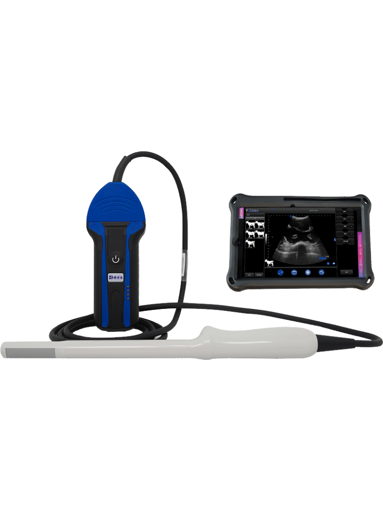
Physical Principles Behind Veterinary Ultrasound Diagnosis
Veterinary ultrasound is one of the most important and widely adopted diagnostic tools in modern animal healthcare. From cattle and pigs to goats and horses, ultrasonography has revolutionized how veterinarians assess internal organs, monitor reproduction, and diagnose various diseases—all without causing harm or stress to the animals. But what exactly makes ultrasound work? What are the physics behind this safe and effective technology?
In this article, we will explore the physical principles of veterinary ultrasound diagnosis in a way that is both informative and easy to understand. We’ll begin with the basics of sound waves, dive into how ultrasound is generated and received, and look at how waves interact with tissues inside the animal’s body. Along the way, we will reflect on how these principles are understood and applied in veterinary practice around the world.

Understanding Ultrasound: The Basics
Ultrasound refers to sound waves with frequencies higher than 20,000 hertz (Hz), which is above the hearing range of humans. While we can hear sounds between 20 Hz and 20,000 Hz, anything above that is considered “ultrasound.” In veterinary diagnostics, ultrasound devices usually operate in a frequency range between 2.0 and 10.0 megahertz (MHz), depending on the species, body part, and depth of tissue being examined.
In essence, ultrasound is a form of mechanical energy. Unlike electromagnetic waves (like light or X-rays), ultrasound needs a medium—such as soft tissue or fluid—to travel through. It cannot travel through air or vacuum effectively. This is why ultrasound gel is applied during scanning: it removes air gaps between the probe and the animal’s skin to allow effective wave transmission.
How Ultrasound Waves Are Created
The creation of ultrasound waves in veterinary devices is based on a fascinating physical effect called the piezoelectric effect. This phenomenon was discovered in 1880 by the Curie brothers in France. The piezoelectric effect allows certain crystals—such as quartz or lead zirconate titanate (PZT)—to convert electrical energy into mechanical energy and vice versa.
Here’s how it works:
-
A veterinary ultrasound machine sends an alternating electrical signal to a special crystal inside the probe (called a transducer).
-
The crystal responds by rapidly vibrating—compressing and expanding along its axis.
-
These rapid vibrations produce sound waves in the ultrasound range. This is known as the “inverse piezoelectric effect,” where electricity becomes sound energy.
-
These waves travel through the animal’s body, encountering tissues of different densities.
How Ultrasound Is Received
Once the ultrasound waves have traveled into the body, they eventually hit boundaries between different tissues—such as between muscle and fat or fluid and bone. At each boundary, some of the ultrasound waves are reflected back toward the probe, while others continue to travel deeper.
When these returning waves (called echoes) strike the same crystal inside the probe, the reverse process happens:
-
The incoming mechanical pressure causes the crystal to vibrate again.
-
These vibrations generate an electric signal in response to the pressure—this is the “direct piezoelectric effect,” where sound energy becomes electricity.
-
The machine then processes these signals into images, waveforms, or sounds.
This two-way conversion of energy is what makes ultrasound such a powerful diagnostic method. The same crystal sends out the signal and listens for echoes—millions of times per second.
How Ultrasound Travels Through the Body
When ultrasound travels through a medium, it behaves like any wave—it can be reflected, absorbed, scattered, or refracted. Understanding these behaviors is crucial for interpreting ultrasound images accurately.
Key concepts include:
-
Reflection: This occurs when ultrasound waves hit a boundary between tissues with different “acoustic impedances.” A strong reflection creates a bright echo on the image (such as from bone or gas).
-
Refraction: Bending of ultrasound waves at an angle when they cross into a medium with different propagation speed.
-
Scattering: Happens when the wave hits small or irregular structures, spreading the energy in different directions. This gives texture to the image.
-
Absorption: Part of the wave’s energy is absorbed by tissue and converted into heat.
-
Attenuation: As ultrasound travels deeper, it loses energy due to absorption and scattering. High-frequency waves attenuate faster than low-frequency ones.
Veterinarians must select the right frequency: higher frequencies give better resolution but cannot penetrate deeply, while lower frequencies reach deeper but with lower image clarity.
Why Acoustic Impedance Matters
One of the most critical factors in ultrasound imaging is “acoustic impedance.” This is a property of a material defined by its density and sound speed. When ultrasound moves from one tissue to another, the change in acoustic impedance determines how much of the wave is reflected or transmitted.
-
Large impedance differences (e.g., between soft tissue and bone) produce strong echoes and appear very bright on the ultrasound image.
-
Small impedance differences (e.g., between similar soft tissues) produce weaker echoes and may appear darker.
This contrast allows veterinarians to distinguish organs, fluids, and abnormalities.
Pulse-Echo Principle in Veterinary Imaging
Most veterinary ultrasound machines use the “pulse-echo” method:
-
The probe emits short bursts (pulses) of ultrasound.
-
After each pulse, the probe waits to receive echoes before sending the next one.
-
The time it takes for the echo to return tells the machine how far away the structure is.
-
The strength of the echo helps determine what type of tissue is being imaged.
In real-time imaging (B-mode or brightness mode), thousands of pulses are sent and received each second, creating dynamic two-dimensional images that veterinarians can interpret on the spot.
Common Imaging Modes in Veterinary Practice
Modern veterinary ultrasound machines typically offer several imaging modes:
-
A-mode (Amplitude mode): Shows echoes as spikes, useful for eye measurements but rarely used elsewhere.
-
B-mode (Brightness mode): The most common, used to create 2D cross-sectional images.
-
M-mode (Motion mode): Captures movement over time, ideal for heart scans.
-
Doppler mode: Measures blood flow speed and direction by detecting frequency shifts in returning echoes.
By combining these modes, veterinarians can not only visualize structures but also assess function—such as heartbeat, fetal movement, or blood circulation.
Global Perspectives: How International Veterinarians Apply the Physics
In Europe, North America, and Australia, ultrasound is an essential part of large animal reproduction programs. Veterinarians routinely scan cattle, sheep, and horses for pregnancy diagnosis, fetal viability, and ovarian assessment using transrectal or transabdominal approaches.
In developing regions, portable ultrasound machines are increasingly used in field conditions. Thanks to advances in miniaturization and battery life, devices are more accessible and easier to use by rural veterinarians and technicians.
Regardless of the country, a solid understanding of the underlying physics improves the quality of ultrasound exams. Proper probe positioning, frequency selection, and image interpretation all depend on the veterinarian’s grasp of wave behavior and tissue interaction.
Limitations and Challenges
While ultrasound is incredibly useful, it has some limitations:
-
It cannot penetrate bone or air-filled structures well (such as lungs or intestines).
-
Image quality depends heavily on operator skill and equipment calibration.
-
Artifacts—such as shadowing or reverberation—can lead to misinterpretation if not properly understood.
Training and experience are therefore essential for making accurate diagnoses.
On-Farm and Clinical Applications
In practice, understanding the physical principles allows veterinarians and livestock producers to make better decisions. For example:
-
Using Doppler ultrasound to assess blood flow in uterine arteries during late pregnancy in mares.
-
Monitoring the reproductive status of dairy cows using real-time B-mode scanning.
-
Assessing muscle and fat distribution in beef cattle for optimal finishing time.
-
Checking for fluid accumulation, abscesses, or cysts in small animals.
As technology improves, so does the resolution and versatility of veterinary ultrasound systems.
Conclusion
Veterinary ultrasound is more than just an imaging tool—it is a product of physics and biology working together to reveal what’s happening inside the animal’s body. From the piezoelectric crystals that generate sound waves to the subtle changes in tissue density that shape the images, every aspect of ultrasound is rooted in fundamental science.
By understanding how veterinary ultrasound works, professionals can use it more effectively and accurately. Whether you’re a veterinarian in France scanning a dairy cow, a technician in Kenya checking goat pregnancies, or a student in China learning the basics of animal diagnostics, mastering these physical principles unlocks a powerful capability in animal health management.
Reference Sources:
-
Kremkau, F. W. (2022). Sonography Principles and Instruments.
-
Nyland, T. G., & Mattoon, J. S. (2014). Small Animal Diagnostic Ultrasound.
-
European Association of Veterinary Diagnostic Imaging (EAVDI) Guidelines.
-
American College of Veterinary Radiology (ACVR) Training Manuals.


