Postpartum Ultrasonic Monitoring of Uterine Wall and Corpus Changes in Ewes
In modern sheep production, managing the postpartum recovery of ewes is critical for ensuring optimal reproductive efficiency and flock productivity. The period following parturition involves a series of complex physiological changes in the uterus, which must return to a non-pregnant state to prepare for subsequent estrous cycles. While palpation and clinical examination offer some insights, veterinary ultrasonography has emerged as a superior, non-invasive technique for monitoring internal reproductive structures in real time.

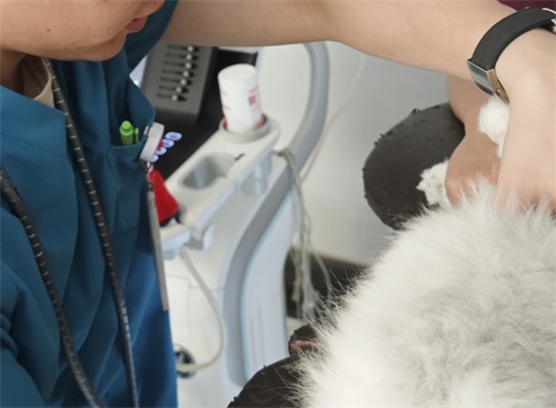
In particular, B-mode veterinary ultrasonography allows producers and veterinarians to visualize the postpartum remodeling of the uterine wall and uterine body. This imaging method has proven valuable not only in research settings but also in routine farm applications, as it helps detect abnormalities, track involution progress, and anticipate future fertility events. This article explores the dynamics of uterine changes in ewes after lambing, as viewed through ultrasound imaging, incorporating both field experience and insights from international studies.
Ultrasound Characteristics of the Uterine Wall in Early Postpartum
From day 1 to day 4 postpartum, ultrasound images typically reveal a thickened uterine wall. This observation is not pathological but reflects the natural mechanical and biochemical aftermath of pregnancy. During gestation, the uterus expands dramatically due to increased content of elongated smooth muscle fibers, collagen, and stromal tissue. Once the fetus is delivered, the uterus immediately begins contracting and shrinking. Without the support of a fetal body and fluid, the uterine wall naturally folds and relaxes.
Furthermore, it is documented that uterine smooth muscle undergoes rapid contraction during the first 48 hours postpartum, with contractions occurring roughly every 2–4 minutes. This leads to a temporary thickening of the uterine wall, as the smooth muscle fibers shorten and overlap. During ultrasound imaging, this appears as a hyperechoic, unevenly thickened structure with a smaller intrauterine cavity. Over the next few days, this thickening gradually subsides.
Uterine Wall Thinning and Involution Progression
By the fifth day postpartum, the uterus begins to show a noticeable decrease in wall thickness on ultrasound. This thinning trend continues steadily, indicating that tissue breakdown and cellular catabolism are progressing. The uterus gradually reverts to its pre-pregnancy state, and by approximately day 22, the ultrasound images show little difference in thickness between the uterine wall and the uterine body. This symmetry marks the completion of uterine involution.
Veterinarians across Europe, North America, and Oceania report similar findings in sheep, goats, and cattle. In clinical settings, ewes scanned regularly post-lambing show this predictable pattern of thickening followed by gradual thinning, which is used as a benchmark for identifying uterine pathologies such as retained fetal membranes, endometritis, or subinvolution.
Hormonal Influences on Uterine Imaging
While uterine involution is a dominant factor in early postpartum changes, hormonal fluctuations also play a role in shaping uterine ultrasound characteristics. Around day 10 postpartum, follicles begin to grow on the ovaries of some ewes, especially in prolific breeds such as the Small-Tailed Han sheep. This follicular development increases estrogen secretion, which in turn promotes endometrial proliferation.
By around day 25 postpartum, studies and on-farm observations have shown that ewes may experience their first ovulation. This hormonal surge can temporarily reverse the thinning trend of the uterine wall, making it appear slightly thickened again under ultrasound. In some cases, this thickening is mistakenly attributed to uterine pathology if not correlated with ovarian activity.
Thus, understanding the endocrinological status of the ewe is essential when interpreting uterine ultrasound results. Many international practitioners recommend concurrent scanning of the ovaries during uterine assessments to identify functional structures like follicles and corpora lutea that may explain imaging anomalies.
Differentiating Normal and Abnormal Postpartum Changes
International veterinary guidelines suggest that the postpartum uterus should show a consistent pattern: initial thickening (days 1–4), gradual thinning (days 5–21), and morphological normalization by day 22–28. Deviations from this pattern may suggest infection, trauma, or delayed involution.
On ultrasound, retained placental tissue appears as echogenic debris within the uterine lumen, while fluid accumulation may appear as anechoic or hypoechoic regions. Thickened uterine walls that do not gradually reduce in size may indicate subclinical inflammation. In such cases, further diagnostics, such as uterine swabs or blood tests, are recommended.
Veterinarians in Australia and the U.S. emphasize the value of regular postpartum ultrasound screenings not only to detect these abnormalities but also to document the natural progression of uterine recovery. This facilitates more targeted treatments and improved reproductive management.
Case Study: Small-Tailed Han Ewe Uterine Monitoring
In a study conducted on Small-Tailed Han sheep, researchers followed uterine involution using daily B-mode ultrasound scans. The study found the uterine wall thickened significantly from day 1 to day 4 postpartum, followed by steady thinning until day 22. By day 25, ovarian scanning revealed the first ovulatory follicle, accompanied by a mild increase in uterine wall thickness due to rising estrogen levels.
The researchers concluded that ultrasound provides accurate, real-time feedback on uterine and ovarian dynamics and could be used to predict when a ewe is ready to rebreed. These findings are consistent with similar research from New Zealand and Germany, where high-frequency linear probes are employed to enhance image resolution in small ruminants.
Technical Considerations in Ultrasound Imaging
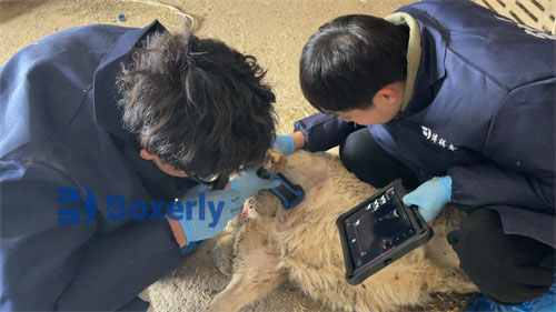
To capture accurate uterine images in ewes, veterinarians typically use a transrectal or transabdominal probe, depending on ewe size and timing postpartum. A 5–7.5 MHz linear or convex transducer provides optimal resolution for visualizing uterine layers and internal contents.
Transrectal ultrasonography, while more invasive, allows higher image resolution and closer contact with uterine structures, especially during the early postpartum period. In contrast, transabdominal scanning becomes more practical as the uterus descends into the abdominal cavity during involution.
Best practices shared by international experts include ensuring adequate probe coupling, using minimal pressure to avoid uterine displacement, and maintaining consistent scanning intervals to track progressive changes.
Practical Applications for Farmers
For sheep farmers and flock managers, incorporating ultrasound monitoring into postpartum protocols can significantly improve reproductive outcomes. Early identification of uterine abnormalities allows for timely intervention, reducing the risk of infertility and prolonged inter-lambing intervals.
Moreover, by observing the completion of uterine involution and the onset of ovarian activity, farmers can make informed decisions about when to introduce rams for natural mating or when to begin estrus synchronization programs. In seasonal breeding systems, this timing is especially critical to maintaining lambing schedules and flock uniformity.
Veterinary ultrasonography also aids in evaluating the success of assisted reproductive techniques, such as artificial insemination and embryo transfer, by confirming uterine preparedness and tracking embryonic development post-conception.
Conclusion
Veterinary ultrasound has transformed the way we observe and understand the postpartum changes in the ewe’s reproductive system. From the initial thickening of the uterine wall due to muscle contraction and tissue remodeling to the gradual thinning during involution and the influence of ovarian hormones, ultrasound provides a non-invasive, real-time window into these physiological events.
As both a diagnostic and management tool, ultrasound imaging empowers veterinarians and sheep producers alike to optimize flock fertility, prevent disease, and reduce economic losses. When interpreted in the context of hormonal cycles and breed-specific traits, ultrasonographic data becomes an indispensable part of modern sheep reproductive health strategies.