Utilizing Animal Ultrasound Scanner to Monitor Postpartum Uterine Recovery in Sheep
For sheep farmers, maintaining reproductive efficiency is the foundation of a profitable flock. Every successful lambing season begins with ensuring that ewes recover well after giving birth. One of the most valuable tools that modern sheep producers and veterinarians have for monitoring the health of postpartum ewes is the animal ultrasound scanner. In this article, I’ll explain how we use ultrasound technology to evaluate postpartum uterine recovery in sheep, why it matters, and how it helps optimize flock management.

Understanding Postpartum Uterine Recovery in Ewes
The postpartum period — also called the puerperium — is a critical stage in a ewe’s reproductive cycle. After lambing, the uterus undergoes involution, a natural process where it shrinks back to its pre-pregnancy size and expels any remaining fluids or tissues. Ideally, this process completes within 3 to 4 weeks, preparing the ewe for her next reproductive cycle. However, factors like difficult births, retained placentas, infections, or poor nutrition can delay uterine recovery and increase the risk of reproductive disorders.
Traditionally, assessing uterine recovery relied on physical examination or waiting for external signs of heat resumption. However, these methods can be subjective and imprecise. With the advancement of veterinary ultrasound, particularly B-mode ultrasonography, we now have a non-invasive, accurate, and real-time method to directly visualize the uterus and assess its condition.
How We Use Ultrasound to Monitor Uterine Recovery
The Basics of B-Mode Ultrasound in Sheep
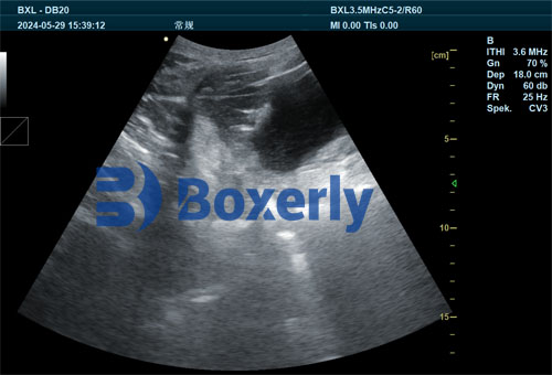
B-mode (brightness mode) ultrasound generates a real-time grayscale image of internal organs. In sheep, we typically conduct transabdominal or transrectal ultrasound examinations, depending on the stage of postpartum recovery and the ewe’s size.
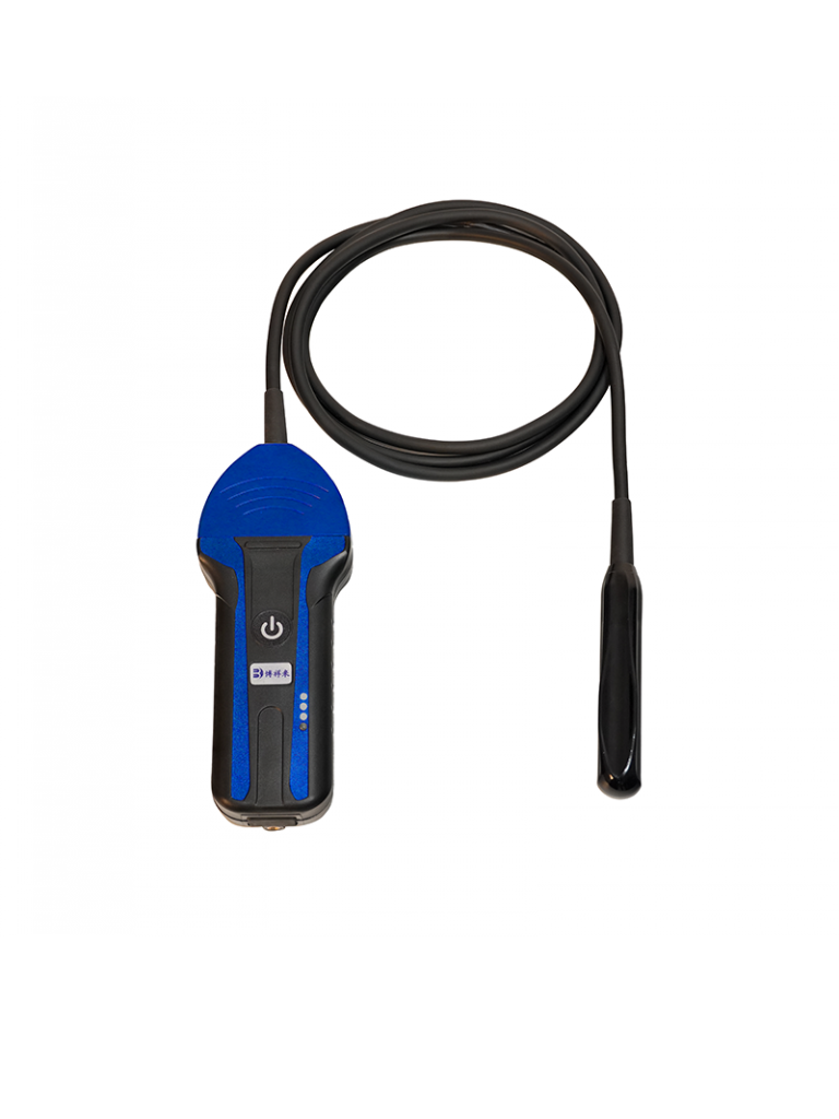
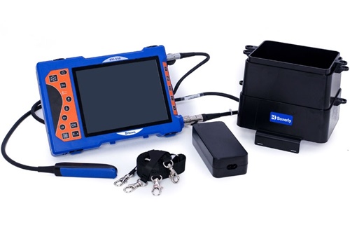
During an examination, the ultrasound probe emits sound waves that bounce off tissues inside the ewe’s body. The returning echoes create a detailed image of the uterus, allowing us to assess its size, shape, and content. With portable scanners designed for farm use, such as the BXL-V50, this process can be done quickly in the field with minimal stress to the animal.
What We Look for During the Scan
-
Uterine Size and Thickness:
A healthy postpartum uterus will steadily decrease in size over the first month after lambing. By measuring the uterine diameter and wall thickness, we can track whether involution is progressing normally. -
Intrauterine Fluid or Debris:
Fluid accumulation or retained tissue appears as hypoechoic (dark) or mixed echogenic structures on the ultrasound image. Detecting these early helps identify cases of endometritis or subclinical infections that might not present obvious external symptoms. -
Endometrial Condition:
The lining of the uterus (endometrium) should return to a smooth, uniform appearance. Thickened or irregular endometrial patterns may suggest inflammation or scarring that could affect future fertility. -
Ovarian Activity:
Although primarily focused on the uterus, we often also scan the ovaries. The presence of developing follicles indicates that the ewe is returning to estrous cycling, which is a good sign of full reproductive recovery.
The Practical Benefits of Ultrasound Monitoring
Early Detection of Postpartum Complications
One of the primary advantages of using ultrasound is the ability to detect issues before they become serious problems. For example, retained fetal membranes or subclinical endometritis can go unnoticed with only visual observation but are easily identified on ultrasound.
In my practice, I’ve seen cases where ewes appeared healthy externally but showed significant intrauterine fluid retention on ultrasound. Early antibiotic treatment based on these findings prevented chronic infections and future reproductive losses.
Non-Invasive and Stress-Free
Unlike some traditional diagnostic methods that may involve manual intrauterine examination, ultrasound is non-invasive and causes minimal discomfort to the ewe. This makes it suitable for repeated examinations over time, allowing for ongoing monitoring without compromising animal welfare.
Making Informed Breeding Decisions
By using ultrasound to assess uterine recovery, we can confidently determine when a ewe is ready for rebreeding. This reduces the risk of breeding ewes with unresolved infections or incomplete involution, which can negatively affect conception rates and embryo survival.

Typical Postpartum Uterine Recovery Timeline on Ultrasound
| Days Postpartum | Ultrasound Findings |
|---|---|
| 1–7 days | Enlarged uterus with some fluid content; thickened endometrium |
| 8–14 days | Gradual reduction in uterine size; decreased fluid volume |
| 15–21 days | Uterine size approaching pre-pregnancy dimensions; minimal or no fluid |
| 22–30 days | Complete uterine involution; clean endometrial lining; possible ovarian follicular activity |
Case Study: Implementing Ultrasound Monitoring on My Farm
Several years ago, I started using the BXL-V50 portable ultrasound scanner as part of our postpartum ewe management protocol. Initially, I was skeptical — after all, our traditional methods seemed “good enough.” But after the first season, the benefits were undeniable.
In one flock of 80 ewes, ultrasound scanning at 14 and 28 days postpartum revealed that about 10% of the ewes had retained fluids or abnormal uterine structures. We treated these animals promptly. The result was a significant improvement in rebreeding rates and a reduction in early embryonic losses during the next breeding season.
Beyond improving reproductive performance, ultrasound scanning gave me peace of mind. I could make breeding decisions based on real data, rather than assumptions.
What International Research Says
In many countries, particularly in Europe, North America, and Australia, veterinarians and researchers have studied the use of ultrasound for monitoring postpartum uterine health in sheep. Key findings include:
-
High Accuracy: Studies show that transrectal ultrasonography can detect uterine abnormalities with sensitivity levels above 90% (Szenci et al., 2018).
-
Economic Benefits: Preventing reproductive failure through early intervention reduces replacement costs and maximizes lifetime productivity (Diskin & Morris, 2008).
-
Welfare-Friendly: Non-invasive monitoring improves animal welfare standards, a growing concern among global consumers (Fisher et al., 2010).
In research led by González-Bulnes (2016), ultrasonography was described as “the gold standard” for non-invasive reproductive monitoring in small ruminants, allowing for both individual animal care and broader herd management optimization.
Integrating Ultrasound into Flock Management
Introducing ultrasound scanning into postpartum care doesn’t require major changes in farm routines. A typical protocol might look like this:
-
Initial Scan (Day 7–10): Detect early signs of retained placenta or fluid accumulation.
-
Follow-Up Scan (Day 21–28): Confirm uterine involution is complete and the ewe is ready for breeding.
-
Pre-Breeding Scan (Breeding Season Start): Evaluate both uterine and ovarian status to optimize breeding strategies.
With portable, durable devices like the BXL-V50, these scans can be done in the field or barn, even by trained farm staff under veterinary guidance.
Limitations and Considerations
While ultrasound is highly effective, it’s not without limitations:
-
Operator Skill: Accurate interpretation requires training and experience.
-
Equipment Cost: While prices have become more accessible, some small-scale farmers may still find the initial investment challenging.
-
Complementary Data: Ultrasound works best when combined with other health indicators, such as body condition scoring, nutrition management, and historical reproductive records.
However, as more producers adopt the technology, training resources and affordable equipment continue to expand globally.
The Future of Ultrasound in Sheep Reproduction
Looking ahead, innovations in veterinary ultrasound technology are poised to make postpartum monitoring even more effective:
-
3D Imaging: New systems can provide detailed volumetric images of the uterus.
-
Artificial Intelligence: Some ultrasound scanners are beginning to integrate AI to assist in detecting abnormalities automatically.
-
Remote Monitoring: Wireless probes and cloud-based data storage allow for real-time veterinary consultation, even in remote locations.
As global livestock production faces increasing pressure to optimize efficiency while maintaining high welfare standards, tools like ultrasound will become essential not just for diagnosing illness but for proactively managing animal health.
Conclusion
The postpartum period is a decisive moment in a ewe’s reproductive life. Using animal ultrasound scanners to monitor uterine recovery offers sheep farmers a safe, accurate, and practical tool to ensure ewes return to full reproductive function quickly and successfully.
By adopting ultrasound into routine postpartum care, producers can improve conception rates, reduce culling, and enhance overall flock productivity. As technology becomes more user-friendly and affordable, I believe ultrasound will become standard practice in sheep management across the world.
Reference Sources:
-
Szenci, O., Takács, E., & Kovács, P. (2018). Use of ultrasonography for postpartum uterine examination in sheep. Small Ruminant Research, 166, 58-64. https://doi.org/10.1016/j.smallrumres.2018.06.005
-
Diskin, M. G., & Morris, D. G. (2008). Embryonic and early foetal losses in cattle and other ruminants. Reproduction in Domestic Animals, 43, 260-267. https://doi.org/10.1111/j.1439-0531.2008.01171.x
-
González-Bulnes, A. (2016). Ultrasonographic imaging in small ruminants reproduction: a review. Veterinary Medicine International, 2016, Article ID 4524829. https://doi.org/10.1155/2016/4524829
-
Fisher, M. W., et al. (2010). Livestock welfare: challenges and solutions. New Zealand Veterinary Journal, 58(3), 119-127. https://doi.org/10.1080/00480169.2010.36884