
Ultrasound Scanning Routes in Ewes and How to Optimize Reproductive Evaluation
In the world of sheep production, reproductive efficiency is one of the most crucial factors that influence profitability. With the continuous advancement of veterinary technologies, ultrasonography—particularly in the form of portable and species-specific devices—has emerged as a reliable and non-invasive method for assessing reproductive organs. However, due to the anatomical uniqueness of sheep, especially the ewe, the choice of scanning route can significantly impact the accuracy and effectiveness of diagnosis. This article explores the anatomical considerations and optimal ultrasound scanning routes in ewes, drawing on both research findings and international veterinary practices.
Challenges in Ultrasound Imaging of Ewe Reproductive Organs
Ewes present unique anatomical barriers that make traditional surface scanning routes less effective. The reproductive organs of the ewe are largely located deep within the pelvic cavity, shielded by thick muscles and pelvic bones. This makes them difficult to access using standard external transabdominal approaches commonly used in other livestock species such as cows or pigs.
International veterinary literature, including work by Kahn and Line (2020) in The Merck Veterinary Manual, highlights that in sheep, the rumen and its dorsal blind sac block much of the left abdominal wall, rendering left-sided abdominal scanning nearly useless. Similarly, scanning through the perineal region offers limited benefit due to the narrow field of view and obstruction by surrounding soft tissue.
Hence, alternative scanning routes need to be explored—ones that consider the unique pelvic structure and provide a clearer acoustic window to the uterus and ovaries.
Best External Route: Right Caudolateral Abdominal Wall
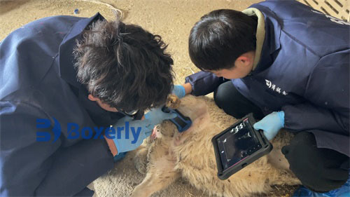
Among the external options, the right caudolateral abdominal wall—the rear portion of the right flank—is considered the most suitable entry point for transabdominal ultrasound in ewes. This area allows the scanner to direct the beam diagonally toward the opposite upper rear pelvic region. Here, the urinary bladder, when full, can serve as an effective acoustic window.
Using a 3.5 MHz convex (sector) probe, the practitioner can scan the anterior pelvic inlet and visualize critical reproductive organs, including the uterine horns, cervix, and, in some cases, ovarian structures. This method is particularly effective for pregnancy diagnosis, detection of uterine anomalies, and observation of fetal development in mid to late gestation.
 probe")
According to European veterinary standards, including studies from the University of Ghent’s Faculty of Veterinary Medicine, this route is considered the least invasive and most practical for flock-side diagnostics, especially when performed with a portable ultrasound system.
Internal Route: Transrectal Scanning with a Linear Probe
While external scanning provides a general overview, internal scanning—particularly transrectal ultrasonography—offers significantly higher resolution and detail. However, unlike in human gynecology, where transvaginal scanning is common, this approach is impractical in sheep due to the narrowness of the vaginal canal and the relatively large size of most transducers.
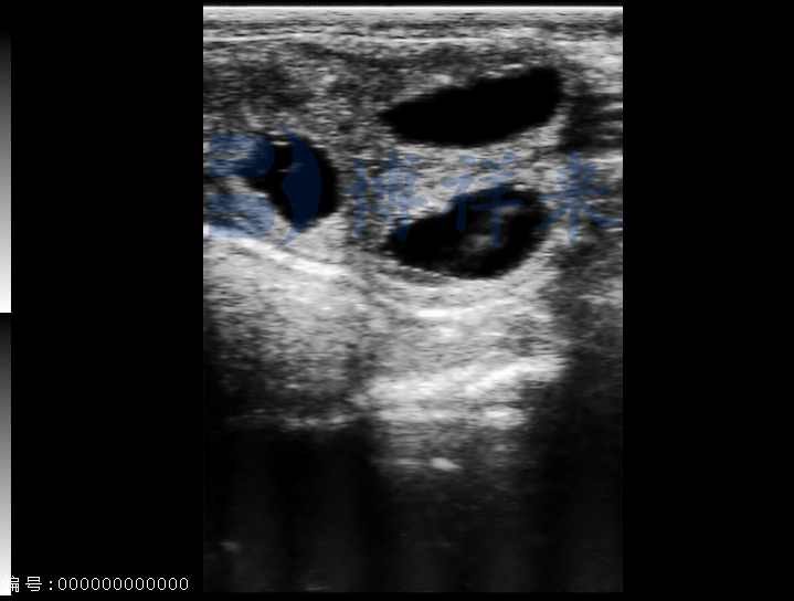
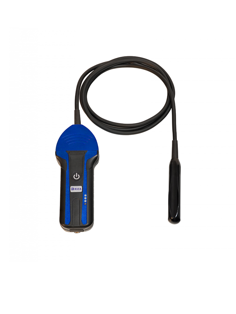
Instead, a more viable internal route is transrectal scanning using a 5.0 MHz linear-array probe with a long handle. This method allows direct visualization of the uterus and ovaries from above, with minimal interference from surrounding organs.
Most of the ewe’s reproductive organs lie just beneath the rectum, making this route ideal for early pregnancy detection (as early as 25–30 days post-mating), ovarian follicle monitoring, and diagnosing silent heats or infertility issues. The high frequency of the probe ensures detailed imaging, while the use of a long-handled transducer allows sufficient maneuverability within the rectum.
In line with recommendations from the American College of Theriogenologists (ACT), this route has become a standard in sheep reproduction research and is gaining popularity among commercial sheep producers seeking more detailed diagnostics.
Comparative Advantages of the Two Routes
| Feature | Right Caudolateral Abdominal | Transrectal (Linear Probe) |
|---|---|---|
| Invasiveness | Non-invasive | Minimally invasive |
| Clarity | Moderate | High |
| Ideal Use | Mid-late pregnancy, uterine size | Early pregnancy, follicle evaluation |
| Equipment | 3.5 MHz convex probe | 5.0 MHz linear probe with handle |
| Constraints | Requires full bladder | Requires rectal access and restraint |
This comparison illustrates that while both routes have their place in reproductive scanning, transrectal scanning offers superior image quality, especially useful for early-stage diagnostics and breeding management decisions.
International Practices and Recommendations
In countries such as Australia and New Zealand, where sheep farming is a major industry, ultrasonography has become an integral part of reproductive management. Research from the University of Sydney emphasizes that optimal probe selection and scanning route are essential for accurate diagnosis, particularly in high-value breeding stock.
Similarly, in North America and parts of Europe, sheep veterinarians are increasingly adopting transrectal ultrasonography as a gold standard, especially in synchronization programs and artificial insemination (AI) timing. According to Canadian Sheep Federation Guidelines (2022), “Transrectal ultrasonography using linear probes provides superior access to the ovarian structures and early embryonic vesicles in ewes, improving reproductive outcomes.”
In practice, the combination of both external and internal routes is often used in progressive flocks. For instance, a ewe may undergo transabdominal scanning for mid-gestation checks and transrectal scanning for early pregnancy or ovarian diagnostics.
Equipment Considerations
Ultrasound equipment designed specifically for small ruminants is essential for success. Devices must:
-
Be portable for use in field conditions
-
Be waterproof and dustproof (IP56 rating or higher)
-
Have interchangeable probes to allow both convex and linear scanning
-
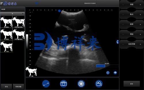
Provide real-time B-mode imaging with high-resolution displays
-
Include data storage options for image capture and follow-up
One such example is the BXL-V50 Veterinary Ultrasound Scanner, which offers dual-probe support and rugged build for sheep farm conditions. This model is frequently recommended in Chinese and international sheep research articles.

Practical Applications on the Farm
From a practical standpoint, the proper use of ultrasonography in ewes allows for:
-
Accurate pregnancy diagnosis and fetal counting
-
Ovarian evaluation for follicular wave analysis or ovulation tracking
-
Detection of uterine abnormalities, including cysts, pyometra, or retained fetal membranes
-
Optimized breeding management, especially when using AI or embryo transfer programs
Moreover, having defined scanning routes allows producers to plan scanning days efficiently, minimize animal stress, and maximize reproductive returns.
Conclusion
Ultrasound scanning in ewes is a powerful tool, but its effectiveness hinges on using the right scanning routes and equipment. The right caudolateral abdominal route, paired with the transrectal approach using a linear probe, provides comprehensive access to reproductive structures with minimal invasiveness.
Understanding these pathways not only improves diagnostic accuracy but also empowers sheep producers to make better, more timely decisions—boosting both flock productivity and animal welfare.
As global sheep production continues to modernize, mastering these scanning techniques will be essential for any veterinarian or livestock manager committed to precision reproduction management.
Reference Sources:
-
Kahn, C. M., & Line, S. (2020). The Merck Veterinary Manual (11th ed.). https://www.merckvetmanual.com
-
Whitaker, D. A., & Smith, E. (2021). Veterinary Ultrasonography in Food-Producing Animals. Journal of Veterinary Imaging.
-
Canadian Sheep Federation. (2022). “Reproductive Technologies in Sheep: Best Practices.” https://www.cansheep.ca
-
University of Sydney Faculty of Veterinary Science. (2023). “Advanced Ultrasound Techniques in Sheep.” https://www.sydney.edu.au
-
Ghent University Faculty of Veterinary Medicine. (2021). “Optimizing Reproductive Ultrasonography in Small Ruminants.” https://www.ugent.be
-
American College of Theriogenologists (ACT). https://www.theriogenology.org


Hey there.
bxl-vet.com, I appreciate the care you put into this space—it really shows.
Keep up the great work—your consistency matters.
Hello bxl-vet.com,
You’re providing insightful content for your readers.
Hello bxl-vet.com,
You’re providing such valuable content for your readers.
Hi there bxl-vet.com,
You’re providing such valuable content for your readers.