Understanding Stillbirths and Mummification in Sows Using Ultrasound Imaging
Modern swine production places heavy emphasis on maximizing reproductive efficiency and litter performance. Among the many challenges swine producers face, stillbirths and fetal mummification remain persistent reproductive issues that lead to economic losses and welfare concerns. Recent research and practical observations suggest that placental efficiency, parity, and sow condition all play interconnected roles in these outcomes. Importantly, the use of veterinary ultrasound technology has enabled swine farmers to monitor reproductive parameters in real-time, aiding in early diagnosis and informed breeding decisions.

In this article, we examine how foreign producers interpret the relationship between stillbirths, mummification, and key reproductive variables such as parity, uterine function, and hormonal balance. We’ll also discuss how ultrasound is used to identify risk factors and improve sow management across various production stages.
The Central Role of Placental Efficiency
One of the leading causes of stillbirths and mummified fetuses is poor placental efficiency. This refers to the placenta’s ability to transfer nutrients and oxygen from the sow to the developing fetus. According to a study published in Theriogenology (Foxcroft et al., 2020), reduced placental efficiency directly correlates with increased fetal mortality in pigs. Inadequate placental development or function can starve fetuses of oxygen and nutrients, causing either late-term stillbirth or intrauterine death followed by mummification.
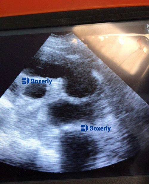
With ultrasound imaging, veterinarians and producers can evaluate the placental and fetal condition during gestation. Hypoechoic or hyperechoic anomalies in the uterine environment, delayed fetal development, or fluid abnormalities may indicate compromised placental performance—alerting caretakers to take precautionary steps such as dietary adjustments or closer monitoring.
Parity’s Impact on Fetal Outcomes
Although placental function is paramount, parity—or the number of litters a sow has produced—is another important factor influencing the likelihood of stillbirths and mummification. Internationally, studies show that parity plays a secondary but significant role in fetal outcomes.
In first-parity (gilts) and older sows (parity ≥6), higher rates of stillbirth and mummified fetuses are commonly reported. In gilts, this is often attributed to an immature reproductive tract and suboptimal progesterone secretion, both of which impact fetal development. In contrast, older sows tend to suffer from uterine fatigue, poor condition, and reduced uterine contractility. These factors prolong farrowing time and compromise fetal oxygenation.
Ultrasound has proven especially useful in identifying these risks. In European farms, for example, veterinary scanners like the BXL-V50 are used to assess fetal size and position in sows of varying parities. High-parity sows often present with larger but fewer fetuses, while gilts may show uneven fetal development. Observing these patterns helps producers determine which animals should be retired or bred selectively.

Farrowing Duration: A Critical Variable
While parity provides a general context for reproductive capability, farrowing duration is a more immediate risk factor for stillbirths. As confirmed by researchers at Iowa State University (Knox et al., 2018), prolonged parturition significantly raises the risk of intrapartum stillbirths, especially in sows beyond their third parity.
Ultrasound evaluation in late gestation can indirectly predict farrowing difficulties by detecting oversized fetuses or malpositioned litters. If such cases are identified early, interventions such as inducing labor or preemptively adjusting sow diets can be implemented.
Age-Related Uterine Involution and Recovery
Every gestational cycle slightly diminishes the uterus’s capacity to fully recover. Repeated pregnancies and deliveries result in microtraumas and inefficient involution—the process by which the uterus returns to its non-pregnant state. Data from studies in Germany (Langendijk et al., 2021) indicate that as parity increases, the uterus takes longer to return to a functional state, thereby reducing the sow’s reproductive performance.
Using real-time B-mode ultrasound, veterinarians can observe uterine tone, wall thickness, and fluid retention during the postpartum period. Poor uterine recovery is often linked to higher incidences of embryonic loss in subsequent pregnancies, an outcome frequently seen in aging sows. Consequently, ultrasound monitoring is not only valuable during gestation but also essential during the weaning-to-estrus interval.
Hormonal Environment and Its Effects on Fetal Viability
Progesterone and estrogen are the two key hormones that govern reproductive success. Progesterone supports the uterine lining and maintains pregnancy, while estrogen prepares the sow for labor and lactation. In gilts, incomplete physiological maturity can result in suboptimal hormone levels, creating a hostile uterine environment for the developing fetus. Additionally, insufficient progesterone production has been linked with embryonic mortality in early gestation.
This hormonal imbalance is especially relevant in young or stressed sows. By combining clinical observation with ultrasonographic scanning, practitioners in the United States and Canada can correlate uterine lining development and corpus luteum function with hormonal adequacy. In cases of insufficient corpus luteum development—visible as small, poorly defined echogenic structures on the ovaries—progesterone supplementation may be recommended.
Immunity and Mummification Risk in Young Sows
Mummified fetuses are more frequently observed in first-parity sows. One major factor is the immature immune response in gilts, making them more susceptible to intrauterine infections that kill fetuses mid-gestation. Viral infections like porcine parvovirus (PPV) or porcine reproductive and respiratory syndrome (PRRS) are often implicated.
Foreign producers increasingly rely on ultrasound to monitor uterine health during critical periods. In Australia, for instance, routine mid-gestation scans help detect signs of abnormal uterine content, such as echogenic debris or irregular fetal heartbeat patterns. Such findings allow for early therapeutic intervention, potentially preventing the progression to fetal mummification.
Statistical Trends and International Insights
Globally, swine data reveal a U-shaped pattern in fetal loss relative to parity: high fetal loss in gilts and old sows, with optimal reproductive outcomes in sows between parity 2 and 5. This is consistent with findings from large-scale studies in Denmark and the Netherlands, where farms use ultrasound-based sow monitoring protocols to optimize breeding schedules.
By analyzing fetal growth, heartbeat, and amniotic fluid volume, foreign producers gather quantifiable indicators of sow reproductive health. This information is then integrated into herd management software, aiding in the early culling of high-risk sows and improving farrowing house outcomes.
The Practical Role of Ultrasound in Decision-Making
On commercial farms, veterinary ultrasound has become a critical tool not just for pregnancy confirmation but for evaluating the quality of that pregnancy. Portable units like the BXL-DZ20 or BXL-V50 offer real-time, high-resolution imaging under farm conditions, making them ideal for regular monitoring.
Some of the most actionable ultrasound parameters include:
-
Fetal heart rate: Indicates viability and stress.
-
Fetal size and number: Helps predict farrowing difficulty.
-
Uterine fluid content: Signals infection or absorption.
-
Ovarian structures: Reflect hormonal status and ovulation timing.
These insights help producers fine-tune breeding schedules, determine sow retention, and prevent reproductive losses.
Conclusion
Stillbirths and mummified fetuses in sows are complex problems rooted in biological and management factors. Placental efficiency, parity, hormonal status, and uterine health all contribute to fetal outcomes, and their effects become more pronounced as the sow ages. By adopting ultrasound technology, producers across the globe have gained a clearer understanding of these reproductive dynamics.
Veterinary ultrasonography allows for real-time, non-invasive evaluation of sow reproductive health. It aids in early diagnosis, prevention, and decision-making—ultimately improving litter outcomes, sow longevity, and farm profitability. As global demand for pork continues to rise, implementing ultrasound in sow herd management is not just beneficial; it’s becoming essential.
References
-
Foxcroft, G. R., et al. (2020). “Placental efficiency and fetal survival in sows: physiological mechanisms and practical implications.” Theriogenology, 150, 93–101. https://doi.org/10.1016/j.theriogenology.2020.01.009
-
Knox, R. V., et al. (2018). “Stillbirths in pigs: Influences of sow and farrowing traits.” Iowa State University Extension. https://www.extension.iastate.edu/
-
Langendijk, P., et al. (2021). “Postpartum uterine health in sows: Implications for fertility.” Reproduction in Domestic Animals, 56(4), 620–628. https://doi.org/10.1111/rda.13901