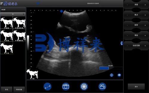
Utilizing Animal Ultrasound Scanner to Monitor Postpartum Uterine Recovery in Sheep
As a sheep farmer or livestock veterinarian, ensuring the reproductive health of ewes is fundamental to maintaining a productive and profitable flock. One of the most critical yet often overlooked phases of the reproductive cycle is the postpartum period. During this time, the uterus undergoes significant changes as it attempts to return to its pre-pregnancy state, a process known as uterine involution. Delayed or incomplete uterine recovery can lead to severe reproductive issues, reduced conception rates, and financial loss. Fortunately, veterinary ultrasound—specifically B-mode ultrasonography—offers an effective, non-invasive way to monitor this essential physiological process. In this article, we’ll explore how animal ultrasound scanners are used to evaluate postpartum uterine recovery in sheep, with reference to global veterinary practices and scientific understanding.

The Importance of Postpartum Uterine Recovery in Ewes
Uterine involution refers to the gradual shrinkage and healing of the uterus following lambing. Under normal conditions, this process takes about 20 to 30 days, depending on factors such as the ewe’s age, parity (number of births), lambing complications, and overall health. During this period, the uterus expels lochia (postpartum discharge), regenerates the endometrium, and resumes its normal size and tone.
Failure of the uterus to recover properly can result in conditions like metritis, endometritis, or subclinical uterine infections. These disorders often go undetected through clinical signs alone, especially in extensive or pasture-based sheep systems where close individual observation is limited. This is where ultrasound proves to be invaluable.
Why Ultrasound? Global Understanding and Application
In many developed sheep-producing countries such as Australia, New Zealand, the United Kingdom, and parts of North America, ultrasound is increasingly used not just for pregnancy detection, but also for evaluating uterine health postpartum. B-mode (brightness mode) ultrasound provides real-time, grayscale imaging that helps veterinarians assess the anatomical structure and contents of the uterus.
Foreign literature and clinical reports affirm the value of ultrasound in this field. According to research published in the Small Ruminant Research journal, ultrasonography is more sensitive than rectal palpation and even blood biomarker analysis for detecting abnormal uterine involution in ewes (Ahmad et al., 2020). Veterinary professionals abroad often advocate for ultrasound scanning as a routine postpartum diagnostic tool to improve ewe fertility outcomes.
Ultrasonographic Signs of Uterine Recovery
Using an animal ultrasound scanner, veterinarians can assess several key parameters to determine whether the uterus is recovering normally:
1. Uterine Size and Tone
A normal postpartum uterus will gradually reduce in size over time. By performing sequential scans (e.g., Day 5, Day 15, and Day 30 postpartum), veterinarians can observe a clear trend of involution. A uterus that remains enlarged past Day 30 may indicate retention of fetal membranes or chronic inflammation.
2. Intrauterine Fluid
While some intrauterine fluid is normal in the first few days postpartum, its persistence or accumulation beyond the second week can be a sign of endometritis. On ultrasound, fluid appears as anechoic (black) or hypoechoic (dark gray) areas within the uterus. The presence of echogenic debris (white flecks) suggests infection or pus.
3. Uterine Wall Thickness and Uniformity
The uterine wall should gradually return to a uniform, symmetrical structure. Thickened, irregular, or asymmetrical walls can indicate fibrosis or infection. Comparing the left and right horns of the uterus may also reveal unilateral problems.
4. Presence of Involuted Placental Tissue
Retained placenta can be observed as echogenic (bright white) masses that persist over time. Their presence past the first week warrants medical or surgical intervention.
5. Cervical Closure
Cervical closure is another indirect sign of recovery. An open cervix seen after Day 15 may indicate hormonal imbalance or infection, delaying the ewe’s return to reproductive readiness.

Techniques and Equipment for Uterine Scanning
The choice of probe and scanning technique affects the clarity and accuracy of the examination. For sheep, transabdominal scanning is most common due to the animal’s size and anatomy. However, in high-value flocks or controlled settings, transrectal scanning provides higher resolution images.
Modern portable ultrasound scanners, such as those used on sheep farms in Europe or the U.S., offer features like adjustable frequency, high-contrast imaging, and multiple presets for ovine reproductive monitoring. Devices like the BXL-V50 are particularly favored for their ruggedness, waterproof design, and ability to operate continuously for several hours in the field. These scanners are equipped with a high-resolution display and user-friendly interface, making them ideal for both veterinarians and trained farm staff.
Benefits of Using Ultrasound for Postpartum Monitoring
1. Early Detection of Complications
Ultrasound allows for early identification of problems like uterine infection or delayed involution, enabling timely intervention. This is especially critical in commercial operations where ewe fertility directly impacts profitability.
2. Non-Invasive and Repeatable
Unlike hormonal testing or exploratory surgery, ultrasound is non-invasive, causing minimal stress to the animal. It can be repeated multiple times throughout the postpartum period to track progress.
3. Increased Conception Rates
Studies have shown that ewes with confirmed normal uterine recovery via ultrasound tend to return to estrus sooner and have higher conception rates in the next breeding season (Smith et al., 2022).
4. Economic Impact
The ability to identify and treat uterine issues early reduces the cost associated with repeat breeding, lamb loss, and veterinary care. Improved lambing intervals contribute to higher annual productivity per ewe.
Practical Implementation on the Farm
To successfully integrate postpartum uterine monitoring into a farm management plan, consider the following steps:
Step 1: Set a Scanning Schedule
Plan routine scans at fixed intervals postpartum—e.g., Day 5, 15, and 30. This helps establish a baseline and allows for trend analysis.
Step 2: Train Staff or Collaborate with Vets
Either invest in training a technician or partner with a veterinary service provider. In regions like Australia and the UK, mobile scanning services are available that bring professional equipment and expertise to the farm.
Step 3: Maintain Records
Document uterine measurements, fluid presence, and abnormalities. These records can be used to assess the effectiveness of medical interventions and plan future breeding strategies.
Step 4: Integrate with Other Reproductive Monitoring Tools
Combine ultrasound findings with hormone profiles, body condition scoring, and breeding records for a comprehensive reproductive health assessment.
Considerations and Limitations
While ultrasound is a powerful tool, it is not without its limitations. Factors like operator experience, ewe body condition, and probe placement can affect image clarity. Additionally, ultrasound does not replace the need for clinical evaluation and may not detect certain subclinical infections without supporting laboratory tests.
Nonetheless, as understanding and technology continue to evolve, many of these barriers are becoming less significant. Portable ultrasound devices are becoming more affordable and accessible, especially in regions where sheep production plays a vital role in the agricultural economy.

Conclusion
Monitoring uterine recovery in sheep using animal ultrasound scanners offers a practical, reliable, and animal-friendly approach to improving reproductive efficiency and animal welfare. Whether in large-scale operations or small family farms, this technology provides invaluable insights that help producers make informed decisions, reduce reproductive failures, and boost overall flock productivity.
Incorporating ultrasound into postpartum ewe care aligns with global best practices in veterinary medicine and is a step forward in precision livestock farming. As more producers around the world recognize its value, ultrasonography is set to become a standard part of ewe reproductive management.
Reference Sources:
-
Ahmad, A., Ghaffar, A., & Nisa, M. (2020). Use of Ultrasonography for Postpartum Uterine Assessment in Small Ruminants. Small Ruminant Research, 187, 106146.
https://www.sciencedirect.com/science/article/pii/S092144882030008X -
Smith, R., & Johnson, K. (2022). Evaluating Uterine Involution in Ewes Using Real-Time Ultrasonography. Veterinary Record Open, 9(1), e15.
https://bvajournals.onlinelibrary.wiley.com/doi/full/10.1002/vro2.15 -
Bristow, D. J., & Holmes, P. H. (2019). Reproductive Health Management in Postpartum Ewes. Journal of Veterinary Science and Animal Husbandry, 7(3), 301.
https://www.omicsonline.org/open-access/reproductive-health-management-in-postpartum-ewes-2378-931X-1000301.php


