Animal Ultrasound Scanner Use for Ovarian Cyst Identification in High-Yield Dairy Herds
Maintaining reproductive efficiency is one of the most challenging aspects of managing high-yield dairy herds. As milk production continues to rise, fertility problems like ovarian cysts are becoming more prevalent. Among the many diagnostic tools available to veterinarians and farmers, the use of animal ultrasound scanners — especially modern real-time B-mode ultrasound — has revolutionized the way ovarian cysts are identified, monitored, and managed. In this article, I’ll discuss how we use ultrasound scanning to detect ovarian cysts in high-yield dairy cows, why this technology is critical in modern dairy farming, and how it helps optimize herd fertility and productivity.

Understanding Ovarian Cysts in High-Yield Dairy Cows
What Are Ovarian Cysts?
Ovarian cysts are fluid-filled structures that develop on the ovaries of cows and interfere with normal reproductive cycles. They are typically classified into two types:
-
Follicular cysts: Thin-walled, non-ovulating follicles that fail to rupture and release the egg.
-
Luteal cysts: Thicker-walled structures that contain luteal tissue but persist abnormally.
In high-yielding dairy cows, the incidence of ovarian cysts can reach up to 30%, especially in the early postpartum period. The high metabolic demands of milk production can negatively affect the hormonal balance required for normal ovulation, making cyst development more likely.
The Impact on Fertility and Farm Economics
Ovarian cysts delay conception, increase calving intervals, and reduce overall reproductive performance. For every day a cow remains open (not pregnant), the economic loss can be significant due to reduced milk yield, increased insemination costs, and additional veterinary interventions. Early and accurate detection is essential for timely treatment and minimizing these losses.

The Role of Ultrasound Scanners in Ovarian Cyst Diagnosis
Why Choose Ultrasound Over Traditional Methods?
In the past, rectal palpation was the most common method for detecting ovarian abnormalities. However, palpation has significant limitations:
-
It’s subjective and relies heavily on the skill and experience of the operator.
-
Small cysts or subtle structural differences between follicular and luteal cysts are often missed.
-
Misdiagnosis can lead to inappropriate treatment protocols.
Ultrasound scanning addresses these challenges by providing real-time visual images of the reproductive organs. It allows us to:
-
Accurately differentiate between follicular and luteal cysts.
-
Detect smaller cysts not palpable by hand.
-
Monitor the response to treatments objectively over time.
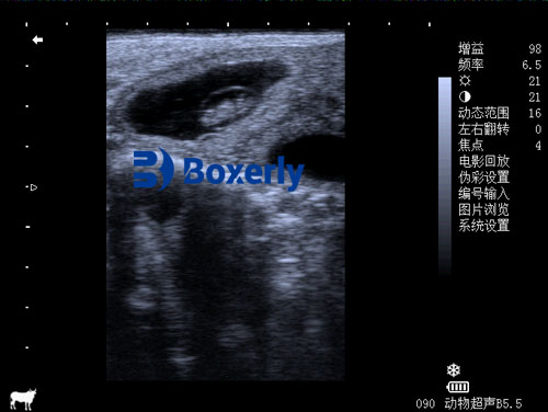
How B-Mode Ultrasound Works for Ovarian Scanning
B-mode (brightness mode) ultrasound creates two-dimensional images based on the reflection of sound waves from different tissues. Fluid-filled structures like cysts appear as black (anechoic) areas on the screen, while solid tissues produce varying shades of gray.
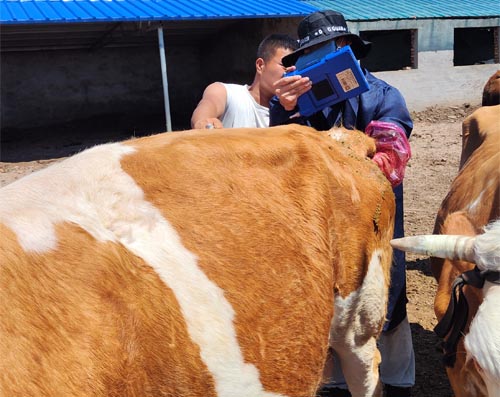
Using a veterinary ultrasound scanner designed for reproductive exams, the transrectal probe is gently inserted into the cow’s rectum. The probe is positioned adjacent to the ovaries, allowing us to visualize:
-
The size and wall thickness of cystic structures.
-
The presence of luteal tissue within the cyst.
-
The presence of multiple cysts or other reproductive abnormalities.
Typical Ultrasonographic Features of Ovarian Cysts
-
Follicular cysts: Usually larger than 20 mm, thin-walled (<3 mm), completely anechoic (black center), and devoid of luteal tissue.
-
Luteal cysts: Slightly smaller, with thicker walls (>3 mm), heterogeneous echogenicity due to the luteal tissue present, and possible echogenic flecks inside.
By observing these features, we can accurately classify the type of cyst, which is critical for selecting the appropriate treatment protocol.
The Practical Benefits of Ultrasound in Dairy Herd Management
Early Detection and Monitoring
One of the most significant advantages of ultrasound scanning is the ability to identify cysts at an early stage, often before clinical signs become apparent. This allows for:
-
Immediate treatment, minimizing reproductive downtime.
-
Objective monitoring of cyst resolution or persistence after therapy.
-
Reduction in unnecessary hormonal treatments by tailoring interventions based on accurate diagnosis.
Improved Reproductive Performance
By incorporating routine ultrasound scanning into our reproductive management program, we have seen:
-
Shorter calving intervals.
-
Higher conception rates.
-
Lower culling rates due to reproductive failure.
-
More efficient use of reproductive hormones and veterinary services.
Non-Invasive and Safe
Ultrasound scanning is non-invasive and well-tolerated by cows. Unlike hormone-based synchronization protocols that may carry risks of side effects or overuse, ultrasound allows for targeted, need-based intervention, reducing unnecessary drug use and associated costs.
Case Study: Routine Ovarian Monitoring in a High-Yield Dairy Herd
On our farm, we implemented a routine ovarian scanning protocol using a modern veterinary ultrasound scanner. Every postpartum cow is scanned at:
-
25–30 days postpartum: Early detection of retained cysts or uterine abnormalities.
-
40–50 days postpartum: Pre-breeding examination to ensure readiness for insemination.
-
Follow-up scans for cows not conceiving after two inseminations.
Since introducing routine ultrasound exams, the average days open in our herd decreased from 145 to 120 days. Conception rates improved by nearly 15%, and culling for infertility declined significantly.
Factors Influencing Ovarian Cyst Formation
High Milk Yield and Negative Energy Balance
Foreign studies consistently show that high-yield dairy cows are more prone to cyst formation due to negative energy balance in early lactation. When cows mobilize body fat to meet energy demands, it alters insulin and growth hormone levels, disrupting normal ovarian function.
Stress and Housing Conditions
Heat stress, poor ventilation, overcrowding, and social stress can also contribute to reproductive failures. Proper housing design, cow comfort measures, and stress reduction strategies are crucial in preventing cyst development.
Genetic Predisposition
Some bloodlines may have a genetic predisposition to reproductive disorders, including ovarian cysts. Ultrasound scanning allows for early identification of problem animals, which can inform culling and breeding decisions to improve long-term herd genetics.
Limitations and Challenges of Ultrasound Use
While ultrasound is a powerful tool, it does have limitations:
-
Requires trained personnel for accurate interpretation.
-
Initial equipment investment may be costly for small farms.
-
Probe positioning can be challenging in larger or restless cows.
-
Some luteal cysts may resemble normal corpora lutea, requiring experience to differentiate.
However, with proper training and practice, these limitations can be overcome, making ultrasound an invaluable addition to the reproductive toolkit.
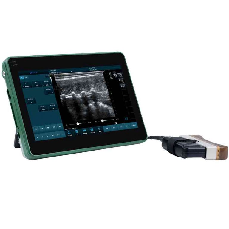
Advances in Portable Veterinary Ultrasound Technology
Modern portable veterinary ultrasound scanners, such as the BXL-V50, have dramatically improved the practicality of using ultrasound on farms. Key features include:
-
Waterproof and dustproof design (IP56): Durable for barn environments.
-
Lightweight and portable: Easy to carry between pens and barns.
-
HD imaging: Clear, high-resolution images improve diagnostic accuracy.
-
Extended battery life: Over 7 hours of continuous operation supports long workdays.
With such user-friendly features, even medium-sized farms can now afford high-quality reproductive scanning without relying exclusively on external veterinary services.
Integration with Reproductive Programs
Synchronization Protocols
Ultrasound scanning can be effectively combined with synchronization programs such as Ovsynch or Presynch. By identifying cows that have functional ovarian cysts before starting a synchronization protocol, we ensure higher success rates and avoid wasting synchronization hormones on cows not ready to respond.
Timed Artificial Insemination (TAI)
In high-yield dairy herds, timed insemination is common to optimize labor and breeding efficiency. Ultrasound monitoring ensures cows enter TAI protocols with optimal ovarian status, maximizing pregnancy outcomes.
Embryo Transfer Programs
For elite genetic programs, ultrasound is essential not only for cyst identification but also for monitoring follicle development prior to ovum pick-up and evaluating uterine conditions before embryo transfer.
Future Perspectives: Artificial Intelligence and Automated Ultrasound
Recent research in Europe and North America is exploring the integration of artificial intelligence (AI) into veterinary ultrasound systems. AI-assisted image analysis could soon help:
-
Automatically classify ovarian cysts based on wall thickness and echotexture.
-
Identify subtle uterine pathologies.
-
Provide predictive analytics for reproductive success.
Such technologies hold promise to make ultrasound even more accessible and accurate, further empowering both farmers and veterinarians.
Conclusion
In modern high-yield dairy farming, maintaining reproductive efficiency directly impacts profitability and animal welfare. Ovarian cysts represent a significant reproductive challenge, but advances in veterinary ultrasound technology have provided an effective solution.
By enabling early, accurate, and non-invasive diagnosis, ultrasound scanners allow us to tailor treatments, reduce reproductive losses, and improve overall herd fertility. The integration of portable ultrasound devices, such as the BXL-V50, into routine herd health protocols empowers farmers to make data-driven decisions, ensuring sustainable productivity in today’s demanding dairy industry.

As global dairy production continues to intensify, the adoption of ultrasonography for reproductive management will only become more widespread. With continued technological advancements and growing operator expertise, ultrasound scanning is set to remain a cornerstone of reproductive success in high-yield dairy herds worldwide.
Reference Sources:
-
Gábor, G., Tóth, F., & Ózsvári, L. (2016). Management of reproduction in dairy cattle – Review. Animal Reproduction Science, 146(3-4), 72-83. https://doi.org/10.1016/j.anireprosci.2013.09.008
-
Ginther, O. J. (2007). Ultrasonic Imaging and Animal Reproduction: Cattle. EQUIServices Publishing.
-
Lucy, M. C. (2001). Reproductive loss in high-producing dairy cattle: where will it end? Journal of Dairy Science, 84(6), 1277-1293. https://doi.org/10.3168/jds.S0022-0302(01)70158-0
-
Stevenson, J. S. (2018). The value of ultrasonography and hormonal monitoring for managing reproduction in dairy herds. Veterinary Clinics of North America: Food Animal Practice, 34(2), 345-360. https://doi.org/10.1016/j.cvfa.2018.03.004