Evaluating Placental Integrity in Fullterm Foal Pregnancies via Doppler Ultrasound
For equine breeders, veterinarians, and farm managers, ensuring the health of both mare and foal during pregnancy is a top priority. One of the most critical periods is the final stage of gestation, when the placenta plays a decisive role in supplying oxygen and nutrients to the rapidly growing foal. Failure to detect placental dysfunction in time can lead to compromised foal health or even pregnancy loss. Fortunately, advances in veterinary ultrasonography, especially Doppler ultrasound, provide a powerful, non-invasive tool to monitor placental integrity in fullterm pregnancies. In this article, I’ll share how we use Doppler ultrasound to evaluate placental health in late-term foals, drawing from both practical experience and global veterinary practices.

Understanding Placental Function in Fullterm Pregnancies
The placenta serves as the essential interface between mare and fetus, enabling gas exchange, nutrient delivery, and waste removal. As gestation progresses, especially in the final trimester, the demands on the placenta intensify. The foal’s weight can double during this period, increasing metabolic requirements and placing more strain on placental circulation.
In fullterm pregnancies, subtle placental insufficiencies may not present obvious clinical signs until problems become severe. Therefore, proactive monitoring is essential. Doppler ultrasound offers real-time insights into placental blood flow, allowing early detection of abnormalities that could jeopardize foal viability.
What Is Doppler Ultrasound and Why It’s Ideal for Placental Evaluation
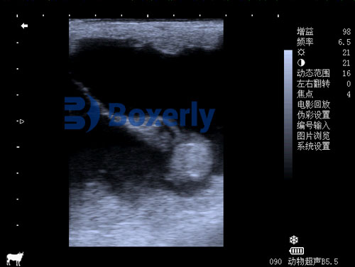
Unlike standard B-mode ultrasound that provides grayscale structural images, Doppler ultrasound measures and visualizes blood flow within tissues and vessels. It detects the frequency shift of sound waves as they bounce off moving red blood cells, converting these shifts into colorful visual maps and quantitative flow data.
Doppler ultrasound is particularly suited for evaluating placental health because:
-
It directly measures uterine and umbilical blood flow.
-
It quantifies vascular resistance, which correlates with placental function.
-
It is entirely non-invasive, safe for both mare and fetus.
Globally, veterinarians are increasingly adopting Doppler ultrasonography as a routine part of advanced equine prenatal care, particularly for high-value sport horse and racing breeds where optimal fetal development is paramount.

Key Placental Parameters Measured by Doppler Ultrasound
When evaluating fullterm equine pregnancies with Doppler ultrasound, veterinarians focus on several crucial indicators:
1. Uterine Artery Blood Flow
The uterine arteries supply blood to the placenta. By placing the Doppler probe transrectally or transabdominally, practitioners can assess peak systolic velocity (PSV), end-diastolic velocity (EDV), and calculate the pulsatility index (PI) and resistance index (RI).
-
A high PI or RI suggests increased resistance, potentially indicating compromised perfusion.
-
A low PI or RI indicates healthy, low-resistance flow, supporting optimal fetal supply.
2. Umbilical Artery Blood Flow
The umbilical arteries carry deoxygenated blood from the fetus to the placenta. Changes in flow velocity, increased resistance, or reversed flow are red flags for fetal distress or placental insufficiency.
3. Middle Cerebral Artery (MCA) Doppler
In some advanced cases, veterinarians may evaluate fetal cerebral blood flow. Brain-sparing effects (redistribution of blood to vital organs) are compensatory signs of chronic hypoxia.
By combining these parameters, veterinarians can build a comprehensive picture of fetal well-being and placental function.
Common Placental Disorders Detectable via Doppler Ultrasound
Several pathologies affecting fullterm foal pregnancies can be identified or monitored with Doppler ultrasound:
Placentitis
One of the most common threats, placentitis (inflammation of the placenta) often results from ascending infections. Doppler ultrasound can detect increased uterine artery resistance and thickened placental tissues, even before external signs like vaginal discharge appear.
Premature Placental Separation (Red Bag)
A dangerous condition where the placenta detaches prematurely before or during delivery. Doppler studies showing abnormal umbilical flow or reduced placental perfusion may signal developing separation.
Fetal Growth Restriction (FGR)
Chronic placental insufficiency limits fetal growth. Doppler assessment can reveal compensatory changes in fetal circulation, prompting early intervention or planned delivery if necessary.
Twin Pregnancies
Twins often struggle for placental territory. Doppler ultrasound helps monitor which fetus may be compromised, informing management decisions.
How We Conduct Doppler Ultrasound on the Farm
On our equine breeding farm, we conduct routine Doppler ultrasound scans during the final 4–6 weeks of gestation. The procedure is simple, well-tolerated by the mares, and yields immediate actionable information.
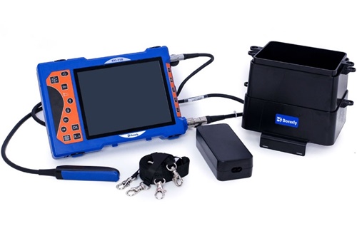
Step 1: Equipment Preparation
We use a high-resolution veterinary Doppler ultrasound machine, ensuring sufficient probe frequency for detailed vascular imaging. A machine like the BXL-V50, equipped with color and pulsed-wave Doppler, offers excellent image clarity and blood flow sensitivity.
Step 2: Probe Placement
Depending on fetal position and operator preference, we may scan transrectally or transabdominally. Transabdominal scanning is often easier in late pregnancy due to uterine enlargement.
Step 3: Vascular Assessment
We target the uterine arteries near the uterine body-placental junction. With careful alignment, we measure flow velocities and calculate indices automatically with the machine’s built-in software.
Step 4: Umbilical Cord Evaluation
If fetal position allows, we visualize the umbilical cord and assess flow characteristics. Any absent or reversed end-diastolic flow is considered an emergency finding.
Step 5: Interpretation and Monitoring
Results are interpreted in the context of the mare’s history, prior scans, and clinical signs. Abnormal findings often prompt increased monitoring, medical intervention (e.g., anti-inflammatory or antimicrobial therapy), or planning for early assisted delivery.
Global Adoption of Doppler Ultrasound in Equine Practice
In countries with advanced equine industries, such as the United States, Australia, Germany, and the United Kingdom, Doppler ultrasonography has become standard for high-risk pregnancies. Research from Colorado State University and University of Liverpool has contributed significantly to developing reference ranges for equine uterine and umbilical Doppler measurements.
In North America, thoroughbred breeding farms routinely employ Doppler imaging to safeguard valuable foals. In Europe, specialized equine reproduction centers integrate Doppler ultrasound into standard late-term pregnancy management protocols.
Even in developing regions, portable machines like the BXL-V50 are expanding accessibility, offering breeders powerful tools once limited to university hospitals.
Benefits of Doppler Ultrasound for Fullterm Foal Pregnancies
Veterinarians and breeders cite several key advantages:
-
Early Detection of Problems: Subclinical placental dysfunction can be identified before clinical deterioration.
-
Guided Medical Management: Therapy can be initiated or adjusted based on real-time data.
-
Reduced Pregnancy Loss: Timely intervention can save both mare and foal lives.
-
Non-invasive and Repeatable: Frequent monitoring causes no harm or stress.
-
Supports Decision-Making: Informs timing of foaling induction, cesarean planning, or intensive neonatal care.
Challenges and Limitations
Despite its many advantages, Doppler ultrasound has certain limitations:
-
Operator Skill Dependency: Accurate measurements require experience and skill.
-
Fetal Positioning: Poor positioning may limit visualization of umbilical vessels.
-
Cost of Equipment: High-quality Doppler machines remain an investment, though increasingly affordable models are entering the market.
The Future of Doppler Ultrasound in Equine Reproduction
As technology advances, newer innovations promise even greater precision. Three-dimensional power Doppler, contrast-enhanced ultrasonography, and artificial intelligence-assisted interpretation are all being researched. These tools could provide even earlier detection of subtle vascular compromise, enhancing fetal-maternal care worldwide.
Moreover, telemedicine is allowing remote specialists to review Doppler scans in real-time, expanding expertise to smaller farms and clinics globally.
Conclusion
In fullterm foal pregnancies, placental health directly influences neonatal survival and long-term foal vigor. Doppler ultrasound empowers veterinarians and breeders with a clear, dynamic view into placental function, transforming pregnancy monitoring from reactive to proactive care.
By routinely evaluating uterine and umbilical blood flow, we can detect issues early, intervene appropriately, and vastly improve outcomes for both mares and foals. As portable, affordable, and user-friendly Doppler ultrasound devices continue to evolve, this invaluable technology is becoming standard across equine industries worldwide, safeguarding the next generation of performance horses.
Reference Sources:
-
Renaudin, C. D., LeBlanc, M. M. (2021). Use of Doppler ultrasonography for evaluation of uterine and umbilical blood flow in the mare. Journal of Equine Veterinary Science, 99, 103405. https://doi.org/10.1016/j.jevs.2021.103405
-
McCue, P. M., Ferris, R. A. (2022). Advances in equine placental evaluation using Doppler ultrasonography. Theriogenology, 180, 58-66. https://doi.org/10.1016/j.theriogenology.2021.12.020
-
Colorado State University Equine Reproduction Laboratory. (2023). Doppler Ultrasound in Equine Pregnancy Management. https://www.csuvth.colostate.edu/erl/
-
Whitaker, D. A., & Smith, E. (2021). Veterinary Ultrasonography in Food-Producing and Performance Animals. Journal of Veterinary Imaging.