
Detection of Abnormal Follicles During Estrus in Female Dogs Using Veterinary Ultrasound Equipment
Monitoring the reproductive health of female dogs is a critical task for breeders, veterinarians, and farm managers. One of the essential aspects of this process is the detection of abnormal follicles during estrus. With advancements in veterinary ultrasound technology, it is now possible to diagnose follicular issues early, allowing for more effective breeding management and health interventions. This article will explore how veterinary ultrasound equipment can be used to detect abnormal follicles during estrus in female dogs, why early detection matters, and what abnormalities to watch for.
Understanding the Estrus Cycle in Female Dogs
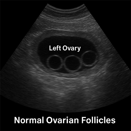
The estrus cycle, often referred to as “heat,” is the period during which a female dog becomes receptive to mating. It typically lasts between 2 to 3 weeks and is divided into four stages: proestrus, estrus, diestrus, and anestrus. Estrus is the key stage for breeding, characterized by ovulation and the presence of mature follicles on the ovaries.
During a normal estrus cycle, follicles grow under the influence of hormones, eventually leading to ovulation. However, abnormalities in follicle development can prevent successful breeding, cause hormonal imbalances, and sometimes result in serious health issues like ovarian cysts or tumors.
The Role of Veterinary Ultrasound in Reproductive Monitoring
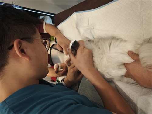
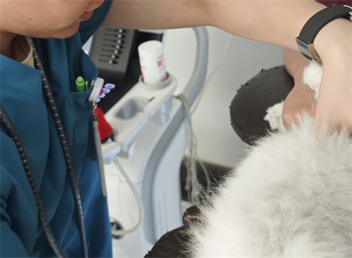
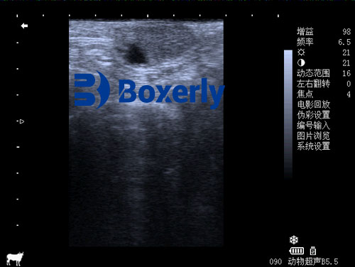
Veterinary ultrasound equipment provides a non-invasive, real-time imaging method for monitoring the reproductive system. High-frequency sound waves create detailed images of the ovaries, uterus, and surrounding tissues, allowing veterinarians to assess follicular development accurately.
Modern ultrasound devices can differentiate between normal and abnormal follicular structures based on size, shape, and internal consistency. They are also valuable for guiding treatment decisions, such as hormonal therapy or surgical intervention.
Identifying Abnormal Follicles Using Ultrasound
There are several types of follicular abnormalities that can be detected via ultrasound:
-
Persistent Follicles: In some cases, follicles fail to ovulate and remain on the ovary. Persistent follicles can continue to secrete estrogen, leading to prolonged estrus symptoms and fertility problems.
-
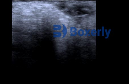
Follicular Cysts: These are fluid-filled sacs that develop from follicles that fail to release an egg. They appear as large, anechoic (dark) structures on ultrasound and can disrupt the normal hormonal cycle.
-
Luteinized Follicular Cysts: Sometimes, a follicle may luteinize (transform into a structure that secretes progesterone) without releasing an egg. These cysts can mimic pregnancy or lead to false hormonal signs.
-
Ovarian Tumors: Though less common, tumors such as granulosa cell tumors can be mistaken for large follicles. They often present as irregular, solid masses on ultrasound and require prompt attention.
Benefits of Early Detection
Detecting abnormal follicles early in the estrus cycle offers several advantages:
-
Improved Breeding Success: Addressing follicular issues promptly increases the chances of successful conception.
-
Health Preservation: Early intervention prevents complications like pyometra (uterine infection), hormonal disorders, and infertility.
-
Cost-Effectiveness: Managing problems early reduces the need for expensive surgical treatments or long-term medication.
Additionally, early diagnosis helps in making informed decisions about whether to proceed with mating or to postpone it until the female dog’s reproductive health is restored.
Best Practices for Ultrasound Examination
To maximize the effectiveness of ultrasound in detecting abnormal follicles, several best practices should be followed:
-
Timing: Perform ultrasound examinations during the early to mid-estrus phase when follicles are most visible.
-
Equipment Selection: Use high-resolution ultrasound machines equipped with linear or microconvex probes for optimal imaging of small structures.
-
Experienced Operators: A skilled veterinarian or trained technician should conduct the scan to correctly interpret the findings.
-
Follow-up Scans: In cases of detected abnormalities, schedule follow-up ultrasounds to monitor changes and response to treatment.
Case Study: Diagnosing a Follicular Cyst
Consider the case of a 4-year-old female German Shepherd presented during estrus for routine breeding assessment. On ultrasound, the veterinarian noted a large, fluid-filled structure on the left ovary, measuring 2.5 cm in diameter—much larger than normal pre-ovulatory follicles. The structure was diagnosed as a follicular cyst.
The dog was treated with hormone therapy to induce ovulation and resolve the cyst. A follow-up scan two weeks later confirmed the resolution of the cyst, and the dog subsequently had a successful breeding season.
This case highlights how early detection and timely intervention can restore reproductive capability and avoid more severe health issues.
Conclusion
Veterinary ultrasound equipment has revolutionized the detection and management of reproductive disorders in female dogs. By identifying abnormal follicles early during estrus, breeders and veterinarians can ensure better reproductive outcomes, safeguard the health of the animals, and make informed management decisions.
For anyone involved in canine breeding or veterinary care, investing in high-quality ultrasound technology and developing expertise in reproductive imaging are essential steps toward improving success rates and animal welfare.


I think this article is really helpful. It explains things clearly and gives practical info for dog breeders. I showed it to my friend Emily—she said it’s super useful too!