Evaluating Fetal Growth in Swine: Application of Real-Time Ultrasound During Gestation
As swine production becomes increasingly data-driven and precision-oriented, monitoring fetal development in sows has emerged as a critical aspect of managing herd health and optimizing productivity. Real-time ultrasonography offers a safe, non-invasive method to assess fetal growth during gestation, allowing veterinarians and producers to make timely, informed decisions. From early pregnancy confirmation to identifying intrauterine growth retardation (IUGR), ultrasound is changing the way producers manage reproductive efficiency and neonatal survival in pig farming.

Understanding Fetal Development in Swine
Swine gestation typically lasts about 114 days and is divided into three roughly equal trimesters. Fetal growth is most rapid during the final third, but critical developmental milestones begin much earlier. Organs such as the heart, liver, and kidneys begin to form as early as day 20 of gestation, with skeletal and muscular development continuing through day 70. From a production standpoint, the ability to assess fetal viability and development during this period can significantly reduce stillbirths, mummified fetuses, and pre-weaning mortality.
Ultrasound plays a vital role in visualizing the internal structures of the uterus, including fetal positioning, fluid volume, heartbeats, and overall size. Unlike palpation or blood-based hormone testing, real-time ultrasonography allows for direct visual assessment of the developing litter—making it an indispensable tool in modern farrowing management.
Real-Time Ultrasound: How It Works in Swine Reproduction
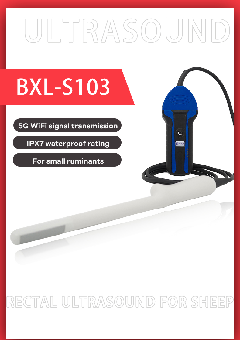
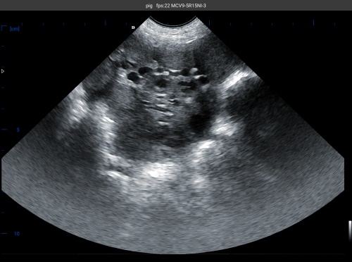
Veterinary ultrasound for swine typically uses B-mode (brightness mode) imaging, which produces 2D grayscale images of internal tissues. A linear or convex transducer probe is applied externally (usually in the inguinal region) or inserted rectally, depending on the sow’s size and stage of pregnancy. Real-time imaging provides instant visual feedback, allowing the operator to detect:
-
The number of developing embryos
-
Fetal heartbeats and movement
-
Amniotic and allantoic fluid volumes
-
Placental thickness and uterine wall condition
-
Relative size of fetuses and potential anomalies
With experience and standardized protocols, operators can measure gestational sac diameter, crown-rump length (CRL), and biparietal diameter (BPD) to estimate gestational age and monitor fetal growth over time. These biometric indicators have been widely validated in both commercial and academic settings.
Key Benefits of Ultrasound in Monitoring Fetal Growth
1. Early Detection of Embryonic Loss
Embryonic loss is most common in the first 30 days of gestation. By performing ultrasound scans between days 21 and 28, producers can identify non-viable embryos early and plan rebreeding strategies, reducing the length of non-productive days.
2. Identification of IUGR and Poor Fetal Development
Intrauterine growth restriction can lead to weak piglets with low birth weights and increased mortality. With ultrasound, abnormal fetal sizes or reduced amniotic fluid volume—early signs of IUGR—can be identified. This helps farmers adjust feeding strategies or provide closer farrowing supervision.
3. Optimized Farrowing Preparation
By confirming viable fetuses and estimating gestational stage, producers can group sows by expected farrowing dates. This supports better planning of labor, facilities, and neonatal care, improving piglet survival and sow recovery.
4. Enhanced Reproductive Record Keeping
Consistent scanning during gestation allows for accurate data collection. Integrated with herd management software, this information supports long-term genetic selection and culling decisions.
Ultrasound Findings Across Gestation Stages
Early Gestation (Days 18–35)
-
Small, anechoic fluid pockets are visible, indicating embryonic vesicles.
-
Embryo viability can be confirmed through the presence of heartbeats.
Mid-Gestation (Days 36–75)
-
Rapid fetal growth is visible. Limb buds, spine, and skull begin to appear.
-
CRL and BPD measurements become more reliable indicators of growth rate.
Late Gestation (Days 76–114)
-
Fetal movement is prominent; internal organs like kidneys and liver are visible.
-
Abnormalities such as fluid imbalance, twisted cords, or deformed limbs may be detected.
-
Size discrepancies between fetuses in the same litter can signal developmental concerns.
Factors Influencing Fetal Growth in Pigs
Several factors affect fetal development, and ultrasound provides insight into how each may be influencing pregnancy outcomes:
1. Sow Nutrition
Undernutrition during early pregnancy can cause embryo loss, while inadequate energy during late gestation may result in underweight piglets. By monitoring fetal size and fluid levels, farmers can adjust nutrient intake at critical times.
2. Parity and Genetics
Younger and older sows tend to have smaller litters or uneven fetal growth. Ultrasound helps track these variations and assists in long-term breeding strategies.
3. Environmental Stressors
Heat stress and high stocking density may impact uterine blood flow and nutrient delivery. Ultrasound can reveal signs of compromised fetal growth, prompting immediate changes in housing or management.
Practical Use in Foreign Farming Systems
In the United States, Canada, and the Netherlands, real-time ultrasonography is increasingly incorporated into standard reproductive protocols. According to studies published by Iowa State University and Wageningen University, farms using ultrasound to monitor gestation reported improved farrowing outcomes, better sow longevity, and reduced neonatal losses.
On U.S. commercial farms, ultrasound is used not just for pregnancy detection but also for weekly monitoring of high-value breeding sows. In Europe, integration with genetic selection programs allows breeders to track fetal growth and link it to specific boar lines or sow family trees.
Advanced portable systems like BXL-V50, equipped with high-resolution imaging and multiple probe options, are helping bridge the gap between veterinary-level diagnostics and practical on-farm application. Devices with long battery life, waterproof design, and intuitive image freezing allow for consistent data capture even in challenging environments.

Comparison with Other Diagnostic Methods
| Method | Accuracy | Invasiveness | Cost | Data Provided |
|---|---|---|---|---|
| Rectal Palpation | Moderate | Moderate | Low | General size, tone |
| Blood Pregnancy Testing | High | Non-invasive | Low | Presence/absence of pregnancy |
| Ultrasound (Real-Time) | Very High | Non-invasive | Moderate | Fetal viability, fluid volume, growth rate, abnormalities |
Ultrasound provides far more detail than traditional methods, making it the best choice for farms aiming for precision pig production.
Economic and Welfare Implications
The use of ultrasound not only improves productivity but also supports better animal welfare. Identifying weak pregnancies or poor fetal growth helps ensure that at-risk sows receive additional care. This reduces stillbirths, improves average weaning weights, and enhances overall herd performance.
Economically, better pregnancy monitoring can improve farrowing rates, reduce feed waste by avoiding unproductive sows, and optimize breeding intervals—each contributing to a more profitable operation.
Limitations and Considerations
Despite its many advantages, real-time ultrasound is not without limitations:
-
Operator dependency: Accurate interpretation requires training and experience.
-
Initial equipment investment: High-quality machines can be costly, though devices like the BXL-V50 offer value through durability and multi-functionality.
-
Limited penetration: In very large or obese sows, image clarity may decrease.
Nonetheless, these limitations are easily outweighed by the benefits in well-managed operations, particularly those with skilled staff or veterinary oversight.
Conclusion
Ultrasound technology has become an essential component in modern swine reproductive management. It enables farmers to go beyond simply detecting pregnancy—offering a real-time window into fetal health, development, and potential complications. By applying this tool throughout gestation, producers can improve neonatal outcomes, reduce sow culling due to reproductive failure, and support a more efficient and welfare-conscious production system.
As precision livestock farming continues to evolve, the role of tools like real-time ultrasound will only grow. Swine producers around the world—whether operating intensive systems in North America or advanced breeding programs in Europe—are turning to ultrasound not just for diagnostics, but for informed, proactive decision-making that benefits animals and business alike.

References:
-
Knox, R. V. (2021). Evaluation of fetal development and pregnancy in swine using ultrasound. Iowa State University Swine Research Reports. https://www.iastate.edu/swine-research/fetal-evaluation-ultrasound
-
Langendijk, P., Soede, N. M., & Kemp, B. (2022). Reproductive performance and fetal growth in modern sows. Wageningen University. https://www.wur.nl/en/show/ultrasound-in-sow-reproduction.htm
-
Whitaker, D. A., & Smith, E. (2021). Veterinary Ultrasonography in Food-Producing Animals. Journal of Veterinary Imaging.
-
National Pork Board. (2023). Precision farming tools in sow management. https://www.pork.org/research/precision-reproduction