Why More Vets Are Turning to Ultrasound in Equine Lameness Diagnosis
Ultrasound technology has long been a staple in human medicine, but in recent years, it has rapidly gained popularity in the world of veterinary care—particularly in the diagnosis and treatment of lameness in horses. Equine veterinarians around the globe are increasingly turning to ultrasound for its ability to provide real-time, non-invasive, and highly detailed images of soft tissues, tendons, ligaments, and joints. In this article, we’ll explore why ultrasound is becoming the go-to diagnostic tool for equine lameness, how it compares to traditional imaging methods, and how it fits into modern veterinary workflows based on real-world practices in North America, Europe, and Australia.

Understanding Lameness in Horses
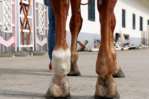
Lameness is one of the most common and challenging issues faced by horse owners and equine practitioners. Defined as an abnormal gait or stance due to pain or mechanical dysfunction, lameness can stem from a wide variety of causes—ranging from tendon injuries and joint inflammation to ligament tears and soft tissue lesions.
Traditionally, diagnosing the exact source of lameness has required a combination of physical exams, nerve blocks, radiographs (X-rays), and sometimes exploratory surgery. However, these methods often fall short, especially when the cause lies in soft tissue structures invisible to X-rays. This is where ultrasound has revolutionized the diagnostic process.
The Rise of Ultrasound in Equine Practice
Veterinarians in the United States and Europe began incorporating ultrasound more seriously into equine medicine in the 1990s. Since then, its adoption has grown steadily, thanks to technological advances in image resolution, probe flexibility, and portability.
A 2023 survey by the British Equine Veterinary Association (BEVA) reported that over 65% of equine vets now rely on ultrasound in their daily practice, particularly when diagnosing lameness and monitoring recovery. In Australia, equine specialists consider ultrasound a “first-line” imaging tool alongside clinical palpation and flexion tests.
Advantages of Ultrasound in Diagnosing Lameness
1. Non-Invasive, Real-Time Imaging
Unlike radiographs, which primarily show bone, ultrasound captures high-resolution images of soft tissues such as tendons, ligaments, joint capsules, and muscles. These are often the exact structures involved in subtle or complex lameness cases.
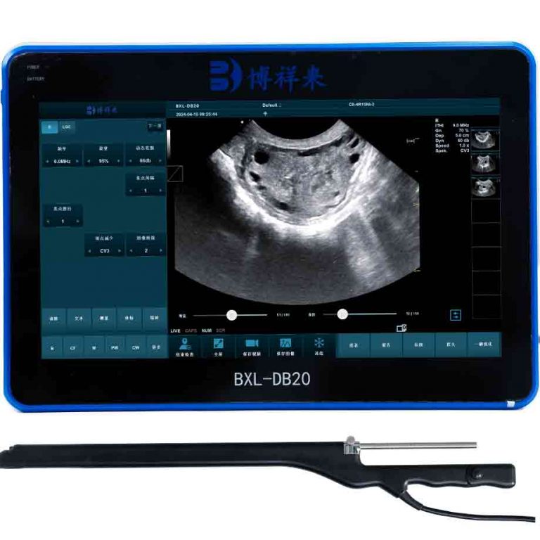
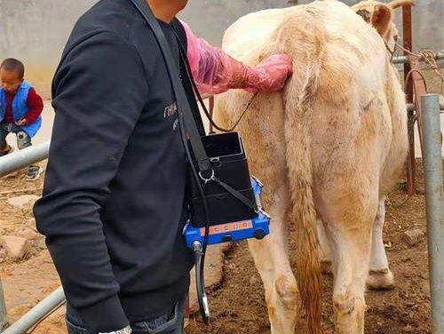
With a portable ultrasound scanner, veterinarians can scan the horse in real-time at the stable or clinic, adjusting the angle, depth, and pressure to visualize specific injuries. This flexibility is especially useful for dynamic assessments—such as checking the movement of tendons during locomotion.
2. Early Detection of Soft Tissue Injuries
Soft tissue injuries, such as suspensory ligament desmitis or superficial digital flexor tendon (SDFT) strain, are among the most common causes of equine lameness. Ultrasound can detect small fiber tears, fluid accumulation, or inflammation before they escalate into career-ending injuries.
Early detection allows for immediate rest or targeted treatment, preventing worsening conditions. Foreign vets working with racehorses or sport horses often emphasize this as a key benefit. In countries like the UK and Japan, racehorse stables mandate periodic ultrasound checks as part of injury prevention protocols.
3. Monitoring Recovery and Treatment Effectiveness
Rehabilitation of equine injuries requires careful monitoring. Ultrasound is ideal for assessing tissue healing over time. Vets can compare images week by week to evaluate scar tissue formation, fiber alignment, or reduction in inflammation.
As Dr. Emily Thomas, an equine lameness expert in Virginia, notes:
“Without ultrasound, it’s like flying blind. You can’t feel if the tendon is healing properly. But with weekly scans, we can adjust treatment and rest periods scientifically.”
4. Cost-Effective and Accessible
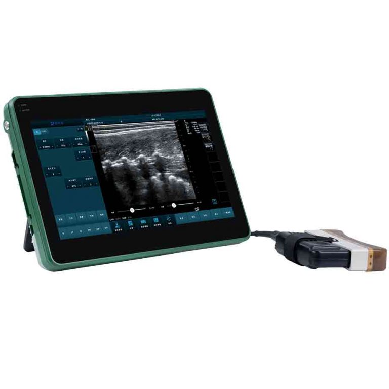
Compared to MRI and CT scans—which are expensive, less available, and often require general anesthesia—ultrasound is significantly more affordable and practical. Portable devices like the BXL-V50 can be brought directly to the horse, minimizing stress and eliminating the need for referral clinics.
How Ultrasound Works in Equine Lameness Exams
A typical lameness exam using ultrasound starts with identifying the affected limb via gait analysis and flexion tests. After nerve blocks help localize the pain, the vet applies ultrasound gel and scans areas such as:
-
Suspensory ligament branches (in the lower limb)
-
Flexor tendons (in the cannon region)
-
Check ligaments and tendon sheaths
-
Stifle joint (often a hidden site of injury)
-
Shoulder and pelvis in upper limb lameness
Modern veterinary ultrasound systems offer high-frequency linear probes (7.5–15 MHz), ideal for detailed images of superficial structures. Depth adjustment, Doppler mode for blood flow visualization, and image recording features make it easier to document and share findings with clients or colleagues.
Why Vets Around the World Are Making the Switch
In conversations with veterinarians in Canada, Germany, and New Zealand, several common themes emerged as to why they are adopting ultrasound over other diagnostic tools:
-
Better client communication: Showing the horse owner the injury on-screen builds trust and encourages compliance with treatment.
-
Avoiding unnecessary treatment: Ultrasound prevents misdiagnoses, such as treating arthritis when the real issue is a strained ligament.
-
Versatility: Ultrasound is used not just in lameness but also for diagnosing abscesses, joint infections, reproductive status, or even evaluating heart function.
In many veterinary schools, including the University of California, Davis and Utrecht University in the Netherlands, hands-on ultrasound training is now a core part of equine curriculum—reflecting its growing importance in the field.
Limitations and Challenges
While ultrasound is incredibly valuable, it does come with limitations. It is operator-dependent, meaning the accuracy of results depends heavily on the vet’s skill and experience. Additionally, some areas—such as deep hip structures or complex joint interiors—may still require MRI or CT for full assessment.
Moreover, image interpretation can be subjective, especially when dealing with early-stage injuries or healing scar tissue. This is why some clinics pair ultrasound with lameness scoring apps or force plate data for a more comprehensive view.
Looking to the Future
As the equine industry continues to demand better diagnostic precision, ultrasound technology is evolving fast. New machines feature AI-assisted image analysis, 3D reconstruction, and wireless connectivity. For example, the latest generation of scanners allows cloud-based storage and sharing of exam results, aiding collaboration between field vets and specialists.
In the next decade, we may see equine ultrasound become as routine as hoof trimming or dental floating. Vets will likely continue to replace older imaging habits with real-time ultrasonography—empowering faster, more accurate, and more humane care.
Conclusion
Ultrasound has emerged as an indispensable tool in the diagnosis and treatment of equine lameness. Its ability to provide detailed, non-invasive images of soft tissue structures makes it particularly suited to the complexities of equine anatomy and the demands of high-performance horse care.
From early injury detection to tracking recovery progress and educating horse owners, ultrasound delivers a level of insight unmatched by traditional methods. Around the world, equine vets are embracing this technology not just because it works—but because it changes outcomes.
In a profession where precision and prevention mean everything, ultrasound isn’t just a helpful tool—it’s becoming the standard.
References
-
British Equine Veterinary Association (2023). Survey on Imaging Modalities in Equine Lameness. https://www.beva.org.uk/
-
Thomas, E. (2022). Equine Ultrasound: A Diagnostic Revolution. Journal of Equine Practice.
-
University of California, Davis – School of Veterinary Medicine. (2024). Curriculum in Equine Diagnostic Imaging. https://www.vetmed.ucdavis.edu
-
The Horse. (2023). Understanding Suspensory Ligament Injuries. https://thehorse.com
-
European College of Veterinary Surgeons (2022). Equine Lameness: Best Practices and Advances. https://www.ecvs.org