Can Veterinary Ultrasound Detect Bladder Stones in Cattle?
For many livestock farmers and veterinarians, managing the health of cattle often centers around reproductive monitoring, fattening programs, and disease prevention. However, one often-overlooked area is the detection and diagnosis of urinary system disorders, particularly bladder stones—also known as urolithiasis. These mineral accumulations can lead to serious complications such as urinary blockage, bladder rupture, or kidney damage, especially in male cattle. But can veterinary ultrasound effectively detect these stones in time to treat or prevent complications?

With growing interest in non-invasive diagnostics, veterinary ultrasound—especially B-mode ultrasonography—is being increasingly recognized for its value in visualizing internal organs, including the bladder and kidneys. In this article, we will explore how veterinary ultrasound works in detecting bladder stones in cattle, share practical experiences from the field, and explain what foreign experts are saying about its effectiveness and limitations.
Understanding Bladder Stones in Cattle
Bladder stones in cattle are usually formed from minerals like struvite (magnesium ammonium phosphate) or calcium carbonate, which can precipitate and accumulate under certain dietary and environmental conditions. These stones can lodge in the urinary tract, particularly in male cattle due to their longer and narrower urethra, leading to potentially life-threatening obstructions.
Common symptoms of bladder stones include:
-
Straining or inability to urinate
-
Abdominal pain or bloating
-
Hematuria (blood in urine)
-
Frequent attempts to urinate with little or no output
Without prompt diagnosis and treatment, the consequences can be severe. Hence, early detection is crucial—and this is where veterinary ultrasound can play a pivotal role.
How Ultrasound Detects Bladder Stones
Veterinary ultrasound, especially B-mode ultrasound, allows real-time imaging of soft tissues and fluid-filled organs. While it cannot “see” bones or mineralized structures as clearly as X-ray, it can still detect highly echogenic (bright) structures within the bladder that cast acoustic shadows—hallmarks of bladder stones.
Ultrasound detection typically involves the following:
-
Scanning Technique: The transducer is placed on the lower abdomen of the animal, often transabdominally in females and small males. In larger bulls or steers, transrectal scanning may be used.
-
Visual Indicators: Stones appear as bright, white structures with clean acoustic shadows beneath them, indicating dense material.
-
Bladder Condition: A distended or thickened bladder wall might indicate irritation or obstruction caused by stones.
-
Real-Time Monitoring: The veterinarian can observe the bladder while manipulating the animal to determine the stone’s size, movement, and location.
Practical Application in Field Conditions
In real-world farm practice, detecting bladder stones with ultrasound offers several advantages:
-
Non-invasive: There’s no need for sedation, surgery, or radiation exposure.
-
Real-time evaluation: This helps in monitoring both the size and mobility of the stones.
-
Quick diagnosis: In emergency cases like suspected urethral obstruction, time is of the essence.
-
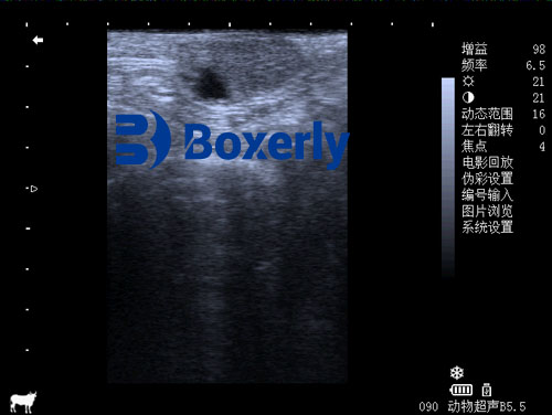
Portable: Modern veterinary ultrasound devices like the BXL-V50 are lightweight, battery-powered, and easy to carry into barns or pastures.
In countries like Australia, Canada, and the U.S., where large herds are managed across vast pastures, field veterinarians often rely on such portable ultrasound systems to detect urinary issues. These scans can guide further intervention—whether medical (e.g., urine acidifiers) or surgical (e.g., urethrostomy or bladder flushing).
Foreign Research and Veterinary Insights
Veterinary literature and studies from around the world support the use of ultrasound for urolithiasis detection in ruminants:
-
USA: According to the Journal of Veterinary Internal Medicine, ultrasound is particularly helpful in diagnosing obstructive urolithiasis in male calves and feedlot steers, where it is often used alongside clinical signs to confirm bladder rupture or stone location.
-
Germany: Veterinary researchers in Berlin have used B-mode ultrasound successfully in diagnosing bladder stones in sheep and goats, recommending its adoption in bovine practice as well.
-
India: In tropical regions where high-phosphorus diets contribute to stone formation, veterinarians frequently use ultrasonography for early-stage detection in breeding bulls.
Dr. Thomas Rausch, a veterinary radiologist in Ontario, remarks, “Ultrasound remains the quickest and most practical method for confirming a diagnosis of urolithiasis in cattle. While not as detailed as a CT scan, it is ideal for real-time, on-farm decisions.”
Limitations and Considerations
Despite its many benefits, ultrasound does have certain limitations:
-
Stone Composition: Some small or non-mineralized stones may not be visible if they do not cast a significant shadow.
-
Operator Skill: Interpretation of ultrasound images requires training. Misdiagnosis is possible without proper experience.
-
Animal Size: In large bulls, the bladder may be difficult to access transabdominally, especially when full of rumen content.
-
Complementary Methods: In some cases, radiography or urinalysis may still be needed for a comprehensive diagnosis.
Yet, these challenges are being steadily addressed through better training, advanced probe technology, and enhanced imaging software.
Case Study: Ultrasound Detection of Urolithiasis in Feedlot Steers
At a cattle operation in Kansas, USA, a feedlot veterinarian used the BXL-V50 portable veterinary ultrasound to investigate a series of steers showing signs of discomfort and reduced urine flow. Upon transabdominal scanning, the team identified multiple bright echogenic masses with posterior acoustic shadows in the bladder, confirming the presence of stones.
Based on the ultrasound results, immediate dietary adjustments and medical treatments were initiated, preventing further obstructions and animal loss. This case highlighted how a portable ultrasound can be lifesaving—not just diagnostic.
Future Perspectives: Advancing the Use of Ultrasound
As technology advances, more accurate and affordable ultrasound machines are being developed specifically for large animal use. Features like enhanced image resolution, Doppler capabilities to assess blood flow, and longer battery life (e.g., 7+ hours in BXL-V50) make these devices more accessible for routine field work.

Moreover, mobile apps and cloud platforms now allow veterinarians to store and analyze ultrasound scans remotely, even sharing them with specialists in other regions. This “tele-veterinary” approach is gaining popularity in North America and Europe.
Additionally, educational programs are increasingly incorporating ultrasound training into veterinary curriculums, ensuring new vets are confident in diagnosing conditions like bladder stones.

Conclusion
So, can veterinary ultrasound detect bladder stones in cattle? The answer is a resounding yes—with some limitations. While not as comprehensive as X-rays or CT imaging, ultrasound provides a highly practical, non-invasive, and cost-effective tool for identifying bladder stones, especially in field conditions.
From small-scale dairy farms to large beef operations, the use of veterinary ultrasound allows for earlier diagnosis, targeted treatment, and better animal welfare. Devices like the BXL-V50 are helping bridge the gap between traditional diagnostic methods and modern, real-time imaging. As more farmers and vets become aware of its value, ultrasound will undoubtedly become a routine part of cattle health management—not just for reproduction or growth, but also for urinary and internal disorders.
References
-
Rausch, T. (2022). “Diagnosis of Urolithiasis in Cattle: Role of Ultrasonography.” Journal of Veterinary Internal Medicine, 36(5), 1123–1130.
https://onlinelibrary.wiley.com/doi/full/10.1111/jvim.16523 -
Prasad, L. N. et al. (2021). “Ultrasonographic evaluation of obstructive urolithiasis in ruminants.” Veterinary World, 14(7), 1842–1847.
https://www.veterinaryworld.org/Vol.14/July-2021/11.pdf -
Heller, M. C. & Braun, U. (2020). “Ultrasonography of the urinary bladder in cattle: Technique and diagnostic utility.” Veterinary Record Case Reports, 8(2), e001101.
https://bvajournals.onlinelibrary.wiley.com/doi/10.1002/vrc2.1101 -
Whitaker, D. A., & Smith, E. (2021). Veterinary Ultrasonography in Food-Producing Animals. Journal of Veterinary Imaging.
https://www.journalvetimaging.org