Veterinary Ultrasound Application in Bovine Ovarian Monitoring
As the global dairy and beef industries become increasingly science-driven, veterinarians and producers alike are embracing non-invasive technologies that offer precision, speed, and efficiency. Among these technologies, veterinary ultrasound stands out as an essential diagnostic and management tool for evaluating the ovaries of cows. Accurate assessment of ovarian structures—including follicles, corpus luteum (CL), and cystic abnormalities—is critical for effective reproductive planning, pregnancy diagnosis, and fertility treatments. From large commercial farms in the United States and Europe to developing herds in Asia and Africa, the application of ultrasound in monitoring bovine ovaries is now seen as a cornerstone of reproductive success.

This article explores the primary ultrasonographic techniques used in bovine ovarian examinations, how these methods are employed in various clinical and field scenarios, and why ultrasound is transforming the way farmers and veterinarians manage dairy and beef cattle reproduction.
Understanding Ovarian Physiology in Cattle
To appreciate the value of veterinary ultrasound, it’s important first to understand the cow’s ovarian cycle. The bovine estrous cycle typically spans about 21 days and involves a coordinated sequence of hormonal and structural changes. During each cycle, a dominant follicle develops, eventually ovulates, and is replaced by the corpus luteum, which secretes progesterone to prepare the uterus for possible pregnancy. In cases where pregnancy does not occur, the CL regresses, and the cycle begins again.
Disruptions in this process—such as follicular cysts, persistent CLs, or silent heats—can cause infertility or delayed breeding, resulting in economic losses due to longer calving intervals and reduced milk production. This is where veterinary ultrasound becomes indispensable.
Primary Ultrasonographic Methods for Ovarian Examination
There are two primary ways to examine the ovaries using veterinary ultrasound in cattle: the transvaginal method and the transrectal method. Both are used globally, although the transrectal approach is more common due to its practicality in the field.
-
Transvaginal Ultrasonography
Transvaginal ultrasonography is a technique in which a specialized probe is carefully inserted into the cow’s vagina. The probe is guided toward the vaginal fornix, just beneath the cervix, and gently manipulated to visualize the uterus and ovaries. This method is especially useful in controlled environments, such as laboratories or reproductive centers, where sedation or restraint can be safely implemented.
Transvaginal ultrasound offers high-resolution images and is often used in advanced procedures like follicular aspiration during in-vitro fertilization (IVF) or oocyte retrieval. However, it is less common on farms due to the need for precise handling and sterility.
-
Transrectal Ultrasonography
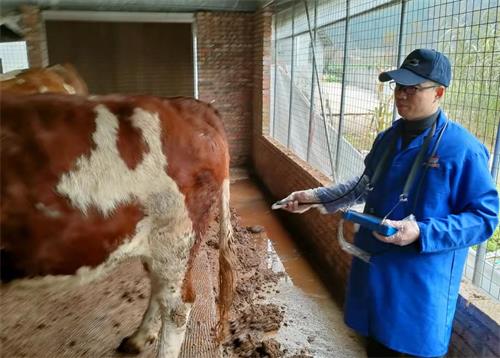
Transrectal ultrasonography is the more commonly used technique on farms around the world. A short, rigid ultrasound probe is inserted into the rectum of the cow, where the operator uses manual palpation to position the probe against the reproductive tract. The rectal wall provides a safe barrier while allowing for detailed imaging of the uterus and ovaries.
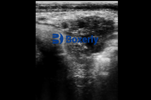
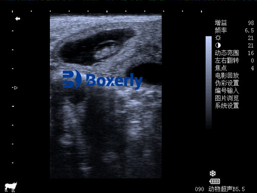
With this method, veterinarians can observe ovarian structures like dominant follicles (often 10–20 mm in diameter), corpus luteum, and small developing follicles. The presence or absence of these structures gives insight into the cow’s reproductive stage and health. Transrectal ultrasonography is non-invasive, causes minimal discomfort to the cow, and can be repeated frequently.
Application in Diagnosing Silent Estrus
One of the most practical uses of ultrasound in bovine reproduction is in identifying cows that are experiencing “silent heat” or silent estrus. These cows go through normal hormonal cycles internally, including ovulation and luteinization, but show little to no external signs of estrus—making them difficult to detect through behavioral observation alone.
According to international studies, approximately 15–25% of postpartum dairy cows may experience silent estrus. This is especially common in high-producing dairy herds, where metabolic stress can interfere with estrus expression. By using ultrasound, veterinarians can identify a corpus luteum on the ovary as early as 28 days postpartum. Progesterone levels tend to rise concurrently, reaching about 3 ng/mL by day 34, indicating the presence of luteal tissue.
However, even with a healthy CL, cows in silent heat may not display mounting or standing behaviors. Ultrasound enables the detection of follicles that fail to ovulate or regress, often persisting without producing a dominant structure. By day 48, progesterone levels may drop to around 1.2 ng/mL, and follicles may remain inactive until a spike in hormone levels around day 72, when visible signs of heat may finally emerge.
With this precise timeline, veterinarians and producers can intervene early, applying hormonal treatments such as prostaglandins or GnRH to synchronize ovulation and improve breeding success.
Detection of Short Luteal Phases
Another reproductive challenge that ultrasound helps manage is the detection of short luteal phases. These are abbreviated cycles in which the corpus luteum regresses prematurely, leading to lower progesterone production and disrupted fertility.
This condition can be observed using transrectal ultrasound between 32 to 72 days postpartum. Typically, cows may show a normal luteal structure and hormone pattern in the first cycle post-calving, but in subsequent cycles, a small corpus luteum may appear briefly and disappear before supporting full uterine preparation.
By regularly monitoring cows with ultrasound, veterinarians can distinguish between a functional and a short-lived CL. Interventions such as adjusted nutrition, improved heat detection protocols, or the use of progesterone-releasing intravaginal devices (PRIDs) can then be introduced to correct the cycle and ensure a successful conception.
Benefits of Using Veterinary Ultrasound in Ovarian Monitoring
Veterinary ultrasound has revolutionized the way modern farms and reproductive specialists manage bovine reproduction. Here are several key benefits seen by practitioners worldwide:
-
Enhanced Reproductive Efficiency
Ultrasound allows for accurate detection of ovulation, corpus luteum formation, and early pregnancy. This enables timely insemination and reduces the number of open (non-pregnant) days in dairy and beef cattle. Shorter calving intervals translate directly into higher milk yields and better herd productivity.
-
Early Diagnosis of Reproductive Disorders
Conditions like follicular cysts, persistent CLs, or ovarian inactivity can be diagnosed early and treated promptly. This reduces the incidence of infertility and helps maintain high conception rates throughout the herd.
-
Precision Breeding and Synchronization
Ultrasound imaging supports the application of synchronization programs by confirming follicular dynamics and corpus luteum development. This makes fixed-time artificial insemination (FTAI) more successful and reduces reliance on labor-intensive heat detection.
-
Non-Invasive and Animal-Friendly
Compared to rectal palpation alone, ultrasound is more precise and causes less discomfort to the animal. It’s also safer for the practitioner, particularly when dealing with larger or nervous cattle.
-
Support for Embryo Transfer Programs
In embryo transfer and IVF protocols, ultrasound is essential for monitoring donor cows, guiding oocyte retrieval, and evaluating recipient cows for suitable uterine conditions. The ability to visualize ovarian activity in real-time increases success rates in these advanced reproductive techniques.
Global Perspective: Adoption and Outlook
In the United States and Canada, veterinary ultrasound is standard practice in commercial dairy operations. Similarly, in European countries like Germany and the Netherlands, it’s integrated into routine herd health visits by veterinarians. In South America, particularly Brazil and Argentina, ultrasound is widely used in beef breeding programs where reproductive intervals are tightly managed.
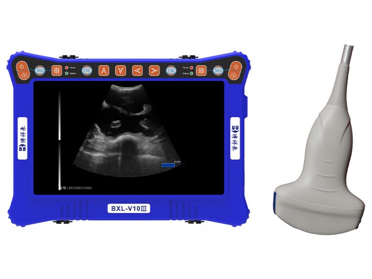
In developing countries, adoption is growing thanks to portable ultrasound units, government extension services, and veterinary training programs. Chinese manufacturers and international companies now produce cost-effective handheld ultrasound machines specifically designed for bovine use.
Looking ahead, technological advancements such as 3D ultrasonography, automated follicle counting, and integration with herd management software will further improve decision-making and herd performance.
Conclusion
Veterinary ultrasound has emerged as an essential tool in monitoring ovarian function in cows. Whether diagnosing silent estrus, managing short luteal phases, or supporting synchronized breeding, ultrasound provides clear, real-time insights into the cow’s reproductive health. Its non-invasive nature, accuracy, and efficiency make it invaluable for farms around the world striving for better fertility, shorter calving intervals, and greater profitability.
As livestock farming continues to modernize, veterinary ultrasound will remain at the forefront of reproductive management, serving not only as a diagnostic device but also as a strategic asset for herd improvement and economic success.
Reference Sources
-
Fricke, P. M. (2020). Reproductive Ultrasonography in Cattle. Veterinary Clinics of North America: Food Animal Practice.
-
Sheldon, I. M., & Dobson, H. (2004). Postpartum uterine health in cattle. Animal Reproduction Science.
-
Lucy, M. C. (2001). Reproductive loss in high-producing dairy cattle: where will it end? Journal of Dairy Science.