Ultrasonographic Examination and Restraint Techniques for the Pig Liver
Ultrasound imaging has become an essential diagnostic tool in modern veterinary practice, especially for livestock management. Among pigs, ultrasonography (commonly known as B-mode ultrasound) is widely employed to evaluate internal organs, particularly the liver, due to its size, complexity, and central role in metabolism and health. As a pig farmer, learning how to perform a proper liver ultrasound and restrain the animal appropriately can significantly improve diagnostic efficiency and reduce stress on the animal. This article explores the practical use of ultrasonography for examining the porcine liver, with a focus on anatomical landmarks, scanning procedures, and best practices for animal restraint.
Anatomy of the Pig Liver in Ultrasound
To perform an accurate liver ultrasound in pigs, it’s crucial to understand the anatomical positioning of the organ. The pig’s liver is more developed compared to other domestic animals like cattle and horses. It is thickest at the center and tapers at the edges. Anatomically, it is located primarily on the right side of the abdominal midline, spanning the costal region and extending toward the xiphoid area of the sternum.
-
Left edge of the liver aligns with the 9th or 10th intercostal space.
-
Right edge typically lies beneath the upper portion of the last intercostal space.
-
Ventral edge may extend 3 to 5 cm past the xiphoid process into the abdominal cavity.
The liver’s convex surface (parietal side) faces the diaphragm and abdominal wall, while the concave surface (visceral side) contacts the stomach and intestines. This anatomical orientation is essential when scanning, as it helps determine the proper transducer placement and angle.
In addition, the gallbladder is situated in the gallbladder fossa of the right medial lobe of the liver. On an ultrasound scan, it appears beneath the 10th or 11th costal cartilage on the pig’s right side and presents as an anechoic (dark) fluid-filled structure, making it relatively easy to identify.
Ultrasound Scanning Procedure
When conducting a liver ultrasound in pigs using a B-mode scanner, the first step is to prepare the scanning area. Proper preparation helps ensure optimal contact between the transducer and skin, enhancing image clarity.
Preparation Steps:
-
Restrain the pig securely (see methods below).
-
Clip or shave the scanning area to remove hair that could obstruct the ultrasound beam.
-
Clean and disinfect the skin surface.
-
Apply coupling gel to ensure effective sound wave transmission.
-
Use a low-frequency transducer (3.5–5 MHz) for better penetration, especially in adult pigs.
The transducer should be held perpendicular to the skin and kept stable to avoid image distortion. Real-time imaging allows the operator to visualize organ margins, blood vessels, and echogenic patterns of hepatic parenchyma.
To capture the best image:
-
Avoid excessive pressure that may compress tissues or distort anatomical structures.
-
Freeze the frame once a clear cross-section is achieved.
-
Record or print the image using an ultrasound printer, video capture system, or digital storage for further evaluation.
Special attention should be paid to minimizing movement, both from the operator and the animal, to avoid motion artifacts during scanning.

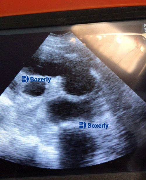
Image Interpretation in Liver Scanning
When interpreting porcine liver ultrasound images, the following should be evaluated:
-
Size and shape of the liver
-
Echotexture (should be homogeneous in a healthy liver)
-
Presence of lesions such as cysts, abscesses, or fatty infiltrates
-
Gallbladder condition (presence of sludge or abnormal size)
A healthy pig liver typically presents as a uniformly echogenic structure with clear margins. Hyperechoic or hypoechoic areas may indicate pathology and should prompt further diagnostic evaluation or lab testing.
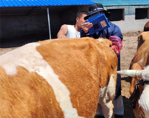
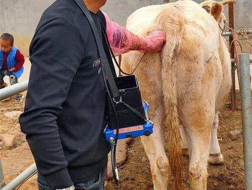
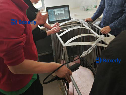
Restraint Methods for Liver Ultrasound in Pigs
Proper animal restraint is vital to ensure both the safety of the operator and the accuracy of the ultrasound exam.
Large Pigs:
-
Restraint in a vertical chute or crate is recommended. This keeps the animal upright, secure, and limits excessive movement.
-
The crate should allow sufficient access to the costal and xiphoid regions for scanning.
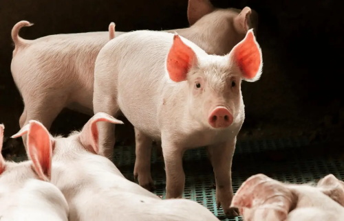
Small Pigs or Piglets:
-
Can be restrained in various positions depending on the scanning site:
-
Standing position if calm and manageable.
-
Dorsal recumbency (lying on the back) on a padded surface if deeper scanning is required.
-
Sitting position may also be used if stable.
-
-
A second handler may be required to gently hold the limbs or secure the head.
In all cases, it’s important to avoid stress or fear in the animal, which can lead to movement and poor-quality images. Gentle handling and a calm environment are key.
Benefits of Ultrasound Liver Evaluation in Pigs
Routine ultrasound examination of the liver in pigs offers several benefits:
-
Early detection of metabolic or infectious liver diseases.
-
Non-invasive monitoring of hepatic conditions without the need for surgical intervention.
-
Evaluation of body condition, especially in breeding sows and boars.
-
Monitoring liver size and gallbladder during antibiotic treatment or diet changes.
-
Guide for liver biopsy, if tissue sampling is necessary for diagnosis.
Additionally, liver evaluation through ultrasonography can support veterinary decisions regarding pig health management, feed adjustments, and culling decisions.
Challenges and Limitations
While ultrasound is highly beneficial, it does come with limitations:
-
Gas in intestines can obscure the liver image.
-
Obesity may reduce image clarity due to increased fat layers.
-
Movement artifacts can distort images if the pig is not properly restrained.
-
Operator experience is essential for accurate interpretation of images, especially in distinguishing between normal and pathological findings.
Despite these limitations, ultrasound remains one of the most effective and practical tools in field diagnostics for pig health.
Conclusion
Ultrasound imaging of the pig liver is a valuable skill for veterinarians and pig farmers alike. By mastering the basic anatomical landmarks, learning how to properly restrain the animal, and developing interpretation skills, one can effectively utilize this technology for early disease detection and overall herd health improvement. As technology advances and portable ultrasound machines become more accessible, integrating ultrasonography into routine farm management will increasingly become the standard for proactive pig care.