
How Is Ultrasound Used for Horses?
Ultrasound imaging, or ultrasonography, is a cornerstone of modern equine veterinary medicine. This non-invasive diagnostic tool employs high-frequency sound waves to produce real-time images of a horse’s internal structures, aiding in the diagnosis and management of various conditions. From reproductive assessments to musculoskeletal evaluations, ultrasonography offers invaluable insights into equine health.
Understanding Equine Ultrasonography
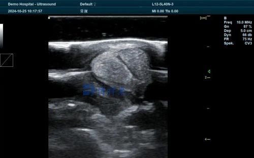
Ultrasonography utilizes sound waves typically ranging from 1.5 to 18 megahertz (MHz) to create images of internal body structures. The most commonly used imaging mode is B-mode (brightness mode) grayscale scanning, which provides two-dimensional images of tissues and organs. A transducer emits sound pulses into the body, and the echoes reflected back are converted into electrical signals to form an image. The quality of the image depends on factors such as tissue density and the frequency of the sound waves used.
Modern ultrasound machines can produce dynamic, real-time images, allowing veterinarians to observe moving structures, such as the beating heart or contracting intestines. These images can be recorded and stored digitally, often in the DICOM (Digital Imaging and Communications in Medicine) format, facilitating easy sharing and analysis.
Reproductive Applications
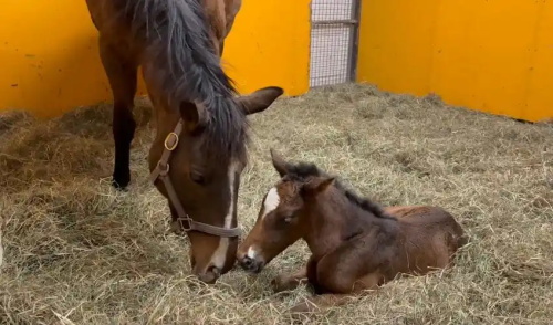
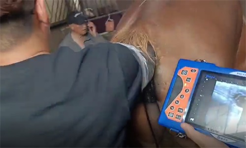
Ultrasound is indispensable in equine reproductive management. For mares, transrectal ultrasonography allows for:Wikipedia
-
Estrous Cycle Monitoring: Assessing follicular development and ovulation timing to optimize breeding schedules.
-
Pregnancy Diagnosis: Detecting pregnancy as early as 14 days post-ovulation and monitoring fetal development throughout gestation.
-
Uterine Health Evaluation: Identifying conditions such as endometritis or uterine cysts that may affect fertility.
In stallions, ultrasonography can evaluate testicular size and structure, aiding in the assessment of reproductive soundness.
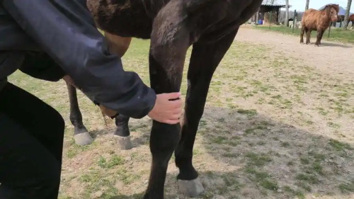
Musculoskeletal Assessments
Ultrasound is a primary modality for diagnosing soft tissue injuries in horses, particularly in the limbs. It enables detailed visualization of:
-
Tendons and Ligaments: Detecting tears, lesions, and inflammation in structures like the superficial digital flexor tendon (SDFT) and suspensory ligament.
-
Muscles: Identifying strains, hematomas, or fibrosis.
-
Joints: Assessing joint capsules, synovial fluid, and cartilage integrity.
Ultrasound also guides therapeutic interventions, such as the precise injection of medications or regenerative therapies into affected areas.
Abdominal and Thoracic Imaging
In cases of colic or other abdominal disturbances, ultrasonography provides critical information about:
-
Intestinal Motility and Position: Identifying obstructions, displacements, or intussusceptions.
-
Organ Evaluation: Assessing the liver, kidneys, spleen, and bladder for abnormalities.
-
Peritoneal Fluid: Detecting abnormal fluid accumulation indicative of peritonitis or hemorrhage.
Thoracic ultrasonography is used to examine the lungs and pleural space, aiding in the diagnosis of pneumonia, pleural effusion, or thoracic masses.

Cardiac Evaluation (Echocardiography)
Echocardiography is the ultrasonographic assessment of the heart, providing insights into:
-
Chamber Size and Wall Thickness: Evaluating for conditions like dilated cardiomyopathy or hypertrophy.
-
Valve Function: Detecting regurgitation or stenosis.
-
Blood Flow Dynamics: Using Doppler ultrasonography to measure flow velocities and detect abnormalities.
This information is vital for diagnosing cardiac diseases and formulating appropriate treatment plans.
Advanced Ultrasonographic Techniques
Recent advancements have expanded the capabilities of equine ultrasonography:
-
Color Doppler Imaging: Visualizes blood flow within vessels and tissues, aiding in the assessment of perfusion and detection of vascular anomalies.
-
Contrast Ultrasonography: Involves the injection of microbubble contrast agents to enhance the visualization of blood flow and tissue vascularity.
-
Ultrasound-Guided Biopsy: Allows for precise sampling of tissues from organs like the liver or kidneys, facilitating accurate diagnosis without the need for invasive surgery.
Limitations and Considerations
While ultrasonography is a powerful diagnostic tool, it has limitations:
-
Bone and Air Interference: Sound waves do not penetrate bone or air effectively, limiting the evaluation of structures obscured by these elements.
-
Operator Dependency: Image acquisition and interpretation require significant skill and experience.
-
Depth Penetration: High-frequency transducers provide better resolution but have limited depth penetration, which may be insufficient for imaging deep structures in large horses.
Conclusion
Ultrasonography has become an integral part of equine veterinary practice, offering real-time, non-invasive insights into a horse’s internal health. Its applications span reproductive management, musculoskeletal diagnostics, internal medicine, and beyond. As technology advances, the precision and utility of ultrasound in equine medicine continue to grow, enhancing the ability of veterinarians to diagnose and treat a wide array of conditions effectively.
References:
-
Esaote VET. “Equine ultrasound: veterinary clinical solutions.” https://www.esaotevet.com/en-US/veterinary-ultrasound/clinical-solutions/equine/
-
IMV Imaging. “An introduction to equine ultrasonography.” https://www.imv-imaging.com/us/academy/an-introduction-to-equine-ultrasonography/
-
Steinbeck Peninsula Equine Clinic. “Ultrasound: An Invaluable Tool In Equine Medicine.” https://www.steinbeckpeninsulaequine.com/post/ultrasound-an-invaluable-tool-in-equine-medicine
-
Lodi Veterinary Care. “Equine Ultrasonography in Equine Practice.” https://www.lodivet.com/equine-ultrasonography-in-equine-practice/
-
Mad Barn. “Ultrasonography for Horses – Equine Research Database.” https://madbarn.com/research-topics/ultrasonography/


Ultrasound’s role in equine care is far more extensive than many realize. It’s impressive how this technology supports everything from reproductive monitoring to diagnosing tendon injuries and heart conditions. The detailed breakdown of how each system is examined helps demystify what happens during a scan. It also makes clear why expertise matters so much—results depend heavily on the operator’s skill.
Интересно, не думал, что у лошадей УЗИ делают почти как у людей. Всё понятно объяснили, даже без спецзнаний заходит. Кто-то реально пробовал такую технику на практике? Работает нормально или просто красиво на словах?
السلام،
أود فهم أفضل عن دور الأشعة فوق الصوتية في رعاية الخيول خاصة في ظروفنا المناخية هنا في السعودية. هل هناك نصائح أو معلومات حول الأجهزة التي تناسب مزارعنا؟
بانتظار ردكم.
I didn’t know ultrasound could pick up on soft tissue injuries so early. Makes me think I should be doing more regular checks, especially during training season. Bookmarked.