Ultrasound Applications in Dairy Cows
As a dairy farmer or veterinarian, understanding the reproductive status and physiological condition of dairy cows is vital for improving herd fertility, reducing breeding failures, and optimizing overall farm productivity. In recent years, the use of veterinary ultrasonography—particularly B-mode ultrasound—has revolutionized cattle reproduction management by providing a real-time, non-invasive, and highly accurate method of diagnosing reproductive and fetal conditions. This article explores the major applications of ultrasound in dairy cows, combining practical experience with current global veterinary insights.

Identifying Corpus Luteum and Follicle Development
Historically, to confirm the presence or stage of development of the corpus luteum (CL) and ovarian follicles, veterinarians had to rely on post-mortem examination of ovaries. This method, while accurate, was impractical for live herd management. Rectal palpation emerged as an alternative but lacked precision—particularly in distinguishing between fluid-filled follicles and cavity-forming CLs, as these structures can feel quite similar.
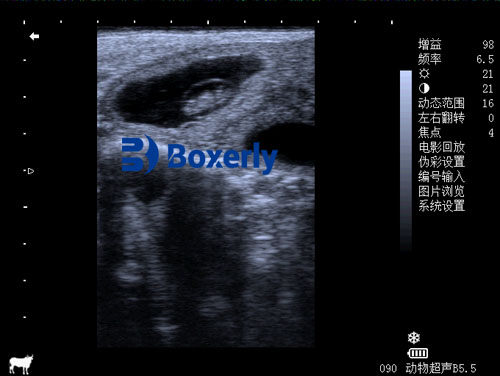
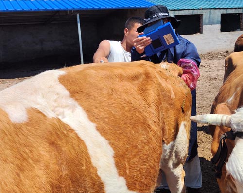
Ultrasound imaging has transformed this process. With a transrectal ultrasound probe, veterinarians can directly visualize the ovary’s surface and internal structure. A developing follicle appears as a round, anechoic (black) area with smooth borders, while the corpus luteum presents as a solid echogenic (gray or white) structure, often with a characteristic internal texture.
The ability to track the precise development stage of the CL is especially important for timing insemination, hormone treatments, or evaluating ovulation success. Reproductive specialists in the U.S. and Europe, such as Fricke et al. (2020), emphasize that synchronized breeding protocols are much more effective when supported by ultrasound-guided follicular monitoring.
Estrus Detection and Timing of Insemination
Accurate estrus detection is a cornerstone of successful breeding. Missed or mistimed insemination leads to extended calving intervals and economic losses. Traditional methods such as observing behavioral signs—mounting, restlessness, or mucus discharge—are often unreliable, especially in high-producing dairy cows where heat signs are subtle.
Ultrasound offers a clearer picture. A mature dominant follicle is easily identifiable, and the presence of estrus mucus in the uterine horns appears as small anechoic patches in the uterine lumen. These indicators, when viewed together, enable precise identification of the estrus stage and the optimal insemination window.
This approach has become standard in countries like Canada and Germany, where dairy farms often rely on routine ultrasound checks to synchronize estrus cycles and plan fixed-time artificial insemination (FTAI) programs.

Fetal Sex Determination
Ultrasound can also be used to determine fetal sex between 55 and 70 days post-insemination. This application is highly valued by dairy producers practicing sexed semen management or strategic culling decisions.
The technique involves inserting a probe into the cow’s rectum and gently scanning the pregnant uterine horn. By identifying fetal orientation and locating the genital tubercle—the tissue that later develops into the penis or clitoris—veterinarians can reliably predict fetal gender. A hyperechoic (bright) genital tubercle located near the umbilical cord indicates a male, whereas its absence or location near the tail suggests a female.
Studies by Curran and Pierson (2022) report an accuracy rate of 90–95% when fetal sexing is performed by skilled technicians using high-resolution ultrasound systems.
Post-Calving Calf Management Support
While not a direct function of the ultrasound itself, the information provided by fetal monitoring helps plan for calving and postnatal care. Once the calf is born, farmers must remove any mucus blocking its airways, disinfect the umbilical cord, and dry the calf to prevent hypothermia.
Ultrasound can contribute to postnatal assessments too. For instance, abnormalities in calf lungs or umbilical structures can be visualized in cases of suspected illness. Ultrasound scans also play a role in detecting subclinical infections or confirming that uterine involution in the dam is proceeding normally.
Western veterinary schools emphasize the integration of ultrasound into broader calf management protocols, advocating early-life health monitoring to reduce calf mortality and improve herd replacement quality.
Monitoring Uterine Health and Postpartum Recovery
Postpartum uterine health directly affects the cow’s readiness to rebreed. Retained fetal membranes, metritis, or delayed uterine involution are common challenges that reduce fertility.
Ultrasound helps detect abnormal uterine contents—such as fluid pockets or thickened uterine walls—allowing for early diagnosis and treatment. For example, dark (anechoic) areas in the uterus post-calving may indicate retained fluids or infection. In Western veterinary practices, these scans are often part of routine postpartum exams conducted 10–20 days after calving.
This method is favored for its speed, safety, and real-time feedback, reducing reliance on systemic antibiotics and improving reproductive outcomes.
Detecting Ovarian Dysfunction and Infertility
Some dairy cows suffer from ovarian inactivity, cystic ovaries, or other reproductive disorders that hinder conception. Palpation alone may miss these issues, but ultrasound can easily identify ovarian cysts—large, round, fluid-filled structures that persist beyond a normal follicular cycle.
By distinguishing between follicular and luteal cysts, veterinarians can tailor treatment (e.g., GnRH or prostaglandin therapies) to restore normal cycling. Reproductive experts in the Netherlands and New Zealand frequently integrate ultrasound monitoring into herd health plans for this reason, reporting improved pregnancy rates and lower culling due to infertility.

Advantages of Using Ultrasound in Dairy Cow Management
The widespread use of ultrasound on dairy farms across the globe is not without reason. Its benefits include:
-
Non-invasive and safe: No pain or distress for the animal.
-
High accuracy: Enables precise identification of reproductive structures.
-
Real-time imaging: Immediate results support fast decision-making.
-
Reduced economic loss: By improving breeding success and reducing open days.
In a study published by the Journal of Dairy Science (Rodríguez-Martínez et al., 2021), ultrasound-based reproductive management was associated with a 15–20% improvement in conception rates when combined with structured breeding programs.
Global Adoption and Future Perspectives
In countries like the United States, Australia, and Denmark, ultrasound-guided reproduction protocols are now considered best practices. Many large-scale dairy operations employ full-time veterinary technicians or train farm staff to use portable ultrasound devices, promoting a culture of precision management.
As ultrasound technology becomes more affordable and compact, smaller farms in developing regions are also adopting its use, supported by agricultural extension services and NGOs.
The future promises even more integration, with AI-enhanced ultrasound systems already being tested to auto-classify ovarian and uterine structures. This will further reduce reliance on expert interpretation and democratize access to reproductive diagnostics.
Conclusion
Ultrasound has become an essential tool in modern dairy cow management. Its ability to non-invasively visualize internal reproductive structures allows for precise timing of insemination, early pregnancy diagnosis, fetal sex determination, and postpartum monitoring. When used consistently, it significantly enhances reproductive performance, reduces economic losses, and improves animal welfare.
On progressive dairy farms across the world, from North America to Western Europe, ultrasound is no longer seen as a luxury but as a vital daily instrument for informed herd management. With continued advancements and accessibility, its role in global dairy production is only expected to grow.
References
-
Fricke, P.M., et al. (2020). “Reproductive management of dairy cows using ultrasound.” Theriogenology, 150, 111–121.
-
Curran, S. & Pierson, R. (2022). “Fetal sex determination in cattle using ultrasonography.” Veterinary Clinics of North America: Food Animal Practice, 38(1), 75–89.
-
Rodríguez-Martínez, H., et al. (2021). “Ultrasound in bovine reproduction: Current practices and future perspectives.” Journal of Dairy Science, 104(5), 5521–5535.
-
Beef Cattle Institute. (2023). “Use of Ultrasound for Growth Evaluation in Cattle.” https://www.beefcattleinstitute.org/ultrasound-growth


