
Will Ultrasound Examinations Be Less Effective If the Pregnant Cow Is Overweight?
Yes—being overweight can significantly reduce the effectiveness of ultrasound examinations in pregnant cows. As a livestock farmer with firsthand experience, I’ve seen how excess fat affects ultrasound imaging. While ultrasound is one of the most reliable tools in veterinary diagnostics, its performance hinges on several physical and biological factors. One of the biggest challenges arises when the subject, in this case a cow, has excessive subcutaneous fat. This article discusses why this happens, how it impacts veterinary ultrasound procedures, and what farmers and veterinarians can do to mitigate it.

How Ultrasound Works in Cattle
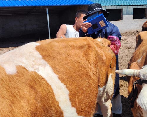
Ultrasound, or ultrasonography, is a diagnostic technique that uses high-frequency sound waves to produce images of internal body structures. In veterinary applications, it is commonly used to assess reproductive organs, monitor pregnancies, and examine soft tissues. The transducer, or probe, emits pulses of ultrasonic energy, which travel through the body and bounce back when they hit tissues of varying densities. These echoes are collected and converted into real-time images.
In bovine practice, transrectal ultrasonography is widely used for reproductive exams. It allows veterinarians to visualize the uterus, ovaries, and developing fetus. However, the clarity and quality of these images are strongly influenced by what the sound waves encounter as they pass through the body.
The Impact of Fat on Ultrasound Imaging
Here is where body condition plays a crucial role. Fat is not just an inert layer—it significantly affects how ultrasound waves travel. Every centimeter of tissue the sound must pass through weakens the signal. So if the cow is overweight, and the ultrasound must penetrate several centimeters of fat before reaching the uterus or fetus, much of the energy will already be absorbed or scattered. This means that by the time the waves reach the organ of interest, there’s not enough remaining energy to produce a clear image.
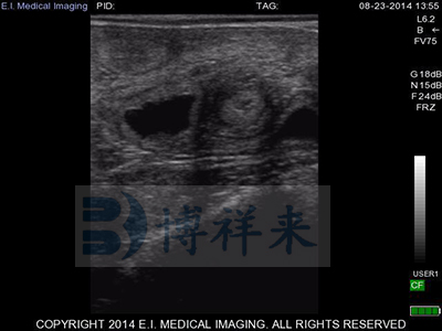
From my experience, scanning an overweight cow often results in blurry, low-contrast images. Sometimes it becomes difficult to distinguish between different structures. In extreme cases, the ultrasound may even fail to detect pregnancy or abnormalities that would otherwise be obvious in a cow of normal weight.
Scientific Reasoning Behind Signal Loss
Ultrasound waves weaken—or attenuate—as they pass through tissue. This loss of signal strength is particularly pronounced in fat because it absorbs and scatters the sound waves more than other tissues. Low-frequency probes can penetrate deeper but provide lower image resolution. High-frequency probes offer better clarity but cannot penetrate thick layers of fat.
In practical terms, using a low-frequency transducer on an overweight cow may help reach the target depth, but the image may lack detail. On the other hand, a high-frequency probe might give up halfway through the fat layer, never reaching the uterus. It’s a trade-off between depth and detail, and excessive fat tips the scale unfavorably.
Real-Life Challenges in the Field
Out in the field, performing ultrasound exams on pregnant cows is not just about the machine—it’s about the cow’s condition. I recall working with a particularly overweight heifer. We had to switch to a lower frequency probe just to get a basic outline of the uterus. Even then, we couldn’t confirm fetal heartbeat without a second scan a week later, after she was put on a lighter feeding regimen and had lost some weight.
These limitations are frustrating for both farmers and veterinarians. Missed or unclear diagnoses lead to poor reproductive management, longer calving intervals, and even missed chances for treating complications early.
What Can Be Done?
To counter these issues, it’s essential to maintain cows at an optimal body condition score (BCS), particularly during breeding and gestation. A BCS of 5 to 6 (on a 9-point scale) is generally ideal for most beef breeds. Not only does this improve fertility, but it also enhances the reliability of ultrasound imaging.
Veterinarians can also adjust scanning techniques:
-
Choose appropriate transducers: Using low-frequency probes for depth and adjusting scanning angles can help.
-
Use more transmission gel: It improves contact between probe and skin, reducing reflection losses.
-
Adjust gain and depth settings: Optimizing the machine’s parameters can partially compensate for weak signals.
-
Early scanning: Imaging earlier in pregnancy—before excessive fat accumulates—can yield clearer results.
Why This Matters to Farmers
Efficient ultrasound exams lead to better herd management. They help in early pregnancy detection, twin diagnosis, and even predicting calving dates. If overweight cows limit the usefulness of ultrasound, then the entire farm operation suffers. Missed pregnancies can mean months of lost productivity. Undiagnosed complications could cost the life of a calf or even the cow.
As a farmer, managing weight is not just about feed costs or aesthetics—it’s about maximizing reproductive efficiency and making the most of every vet visit.

Conclusion
So, will ultrasound examinations be less effective if the pregnant cow is overweight? Absolutely. The physics of ultrasound means that fat acts like a sponge, absorbing the sound waves before they can do their job. This results in weaker signals and poor images, which may hinder accurate diagnosis.
But the good news is, with proactive body condition management and smart scanning techniques, these limitations can be minimized. By understanding how weight affects ultrasound imaging, we as farmers can make informed decisions that benefit both the animals and the bottom line.
References
-
Merck Veterinary Manual – Ultrasonography in Large Animals
-
University of Minnesota Extension – Body Condition Scoring Beef Cows
-
American Association of Bovine Practitioners – Reproductive Ultrasonography


I found this article really interesting because it talks about a problem we often overlook. It made me realize how body condition can affect ultrasound results. I think this kind of information is very helpful, especially for people working with cattle like me.