Veterinary Ultrasound Role Observing Sow Follicle Development
In modern pig farming, optimizing sow reproductive performance is essential to improving productivity and profitability. One of the most critical factors influencing sow fertility is the growth and development of ovarian follicles, which govern the estrous cycle and ovulation timing. Veterinary ultrasound has emerged as an indispensable tool for monitoring follicle development in sows, enabling producers and veterinarians to make informed management decisions that enhance reproductive efficiency.

This article explores how veterinary ultrasound is applied in observing sow follicle development, emphasizing the influence of boar exposure on follicular growth, the physiological significance of follicles during estrus, and practical implications for breeding management. We also incorporate perspectives from international research and pig production systems to provide a comprehensive understanding of this technology’s impact.
Understanding Follicle Development in Sows
The sow’s reproductive cycle centers on the development of follicles within the ovaries. Follicles are small fluid-filled sacs that contain oocytes (egg cells). Their growth is crucial because mature follicles release viable oocytes for fertilization during ovulation. The follicular phase precedes ovulation and is marked by rapid follicle growth and increased estrogen secretion, which triggers estrous behavior.
Follicle development is regulated by complex neuroendocrine signals along the hypothalamic-pituitary-gonadal axis. Follicle-stimulating hormone (FSH) stimulates the growth of several follicles, but usually only one or a few reach full maturity and ovulate. The size and number of follicles at any given time influence the timing and expression of estrus, as well as subsequent fertility.
Boar Exposure and Its Influence on Sow Follicle Development
A key factor accelerating follicular growth and estrus onset is the exposure of gilts and sows to mature boars. International research consistently demonstrates that boar presence exerts a strong stimulatory effect on the reproductive physiology of female pigs.
Boars produce specific pheromones through their submaxillary and preputial glands, which act as external hormonal stimuli. These pheromones trigger neuroendocrine responses in gilts, accelerating ovarian follicle development and advancing the age at first estrus. Behavioral cues such as the boar’s mounting attempts and physical presence further enhance this effect.
In practice, gilts typically experience their first estrus between 170 and 260 days if unexposed to boars. However, contact with mature boars during the critical period of 150 to 170 days significantly reduces the age at puberty. Studies show that introducing a mature boar to a group of gilts results in a marked estrus peak 5 to 7 days later, advancing puberty by 30 to 40 days compared to unexposed groups.
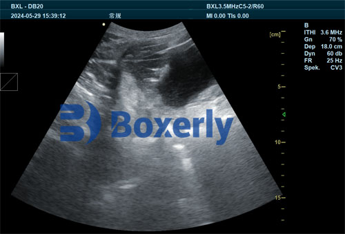
Veterinary ultrasound enables direct observation of these changes by imaging the early development of follicles in gilts exposed to boars. Follicles tend to grow earlier and larger under boar influence, which can be detected as increased follicular size and number on ultrasound scans. This capability allows producers to confirm the effectiveness of boar exposure and adjust management accordingly.
Furthermore, boar contact after weaning can shorten the weaning-to-estrus interval in sows, improving reproductive turnover rates. Monitoring follicle development with veterinary ultrasound during this period helps identify sows that respond well to boar stimulation and those that may require additional interventions.

Veterinary Ultrasound Monitoring Follicle Development and Estrus Expression
Veterinary ultrasound provides a non-invasive, real-time window into the ovary’s dynamic follicular changes, making it an invaluable tool for predicting estrus and timing insemination.
During estrus, follicles undergo rapid enlargement, with the largest follicles reaching diameters typically between 6 and 10 mm before ovulation. Ultrasound can detect these follicles’ presence and growth rate, correlating closely with the sow’s behavioral signs of heat.
Estrogen produced by the granulosa cells within growing follicles induces estrous behavior, including restlessness, vulvar swelling, and acceptance of the boar. It also prepares the reproductive tract for sperm transport and embryo implantation by promoting uterine lining growth.
By visualizing follicle size and number on veterinary ultrasound, producers can better identify optimal breeding windows, enhancing conception rates and litter sizes. Ultrasound assessment of follicles also helps distinguish sows that may be anestrous or have delayed follicular development, enabling early remedial action.
Impact of Follicle Size on Post-Weaning Reproductive Performance
One of the most critical reproductive management challenges is minimizing the interval from weaning to estrus. This interval largely depends on follicle development at weaning.
Studies reveal considerable variability in follicle numbers and sizes in sows at weaning. Sows with numerous small follicles at weaning generally experience longer delays before exhibiting estrus and ovulation, as these follicles require more time to mature.
Conversely, sows presenting medium to large follicles shortly after weaning tend to resume estrous cycles more quickly. Veterinary ultrasound allows precise measurement of follicle size distributions, serving as a predictor for the weaning-to-estrus interval.
This insight empowers farm managers to implement nutritional or hormonal interventions targeting sows with delayed follicle development, potentially reducing non-productive days and improving herd fertility.
International Perspectives and Applications
Globally, pig producers and veterinarians have increasingly embraced veterinary ultrasound to optimize reproductive management. In countries with intensive pig production such as the United States, Denmark, and Spain, routine ultrasound monitoring of sows’ ovarian follicles is becoming a standard practice.
Research from these regions supports the effectiveness of boar exposure combined with ultrasound monitoring in advancing puberty in gilts and shortening weaning-to-estrus intervals in sows. Advanced ultrasound machines with higher resolution enable detailed imaging of follicle morphology and size distribution, facilitating precision breeding.
In some European farms, ultrasound data is integrated with computerized herd management systems, allowing real-time tracking of each sow’s reproductive status and tailoring interventions at the individual level. This approach exemplifies the future of precision livestock farming.
Advantages of Veterinary Ultrasound in Sow Follicle Monitoring
-
Non-invasive and stress-free: Ultrasound does not harm animals and can be performed repeatedly without adverse effects.
-
Real-time visualization: Immediate feedback on follicle size and number aids prompt decision-making.
-
Improved breeding accuracy: Accurate timing of insemination based on follicle development enhances conception rates.
-
Early detection of reproductive disorders: Ultrasound can reveal cystic follicles or anestrus conditions that require veterinary treatment.
-
Facilitates research and genetic improvement: Monitoring follicle development supports selection of sows with superior reproductive traits.
Practical Recommendations for Pig Producers
To fully leverage veterinary ultrasound’s benefits in sow reproductive management, producers should consider the following:
-
Implement controlled boar exposure: Introduce mature boars to gilts between 150 and 170 days of age to stimulate early follicular development.
-
Schedule ultrasound scans strategically: Perform follicle assessments pre-puberty, post-weaning, and pre-breeding to monitor follicular dynamics.
-
Train farm personnel and veterinarians: Skilled operation of ultrasound equipment is essential for accurate interpretation of images.
-
Combine ultrasound data with behavioral observations: Follicle size should be correlated with estrous signs to optimize insemination timing.
-
Integrate reproductive monitoring into herd management software: Digital records improve tracking and decision support.
Conclusion
Veterinary ultrasound has revolutionized the understanding and management of sow follicle development in pig production. Its ability to non-invasively visualize ovarian follicles offers unparalleled insight into reproductive physiology, enabling producers to enhance estrus detection, advance puberty onset, shorten weaning-to-estrus intervals, and ultimately increase reproductive efficiency.
Boar exposure remains a powerful natural stimulus that accelerates follicle growth and estrus expression, effects that are clearly observable and quantifiable with veterinary ultrasound. The combination of these management practices supported by ultrasound technology aligns with global trends in precision livestock farming, promising improved sow fertility and farm profitability.
As international experience demonstrates, veterinary ultrasound is not just a diagnostic tool but a critical component of proactive reproductive management in modern pig farms. Its adoption worldwide continues to grow, driven by the demand for higher productivity, better animal welfare, and sustainable pork production.


