Equine Tendon Injuries: Diagnosis and Management
As a horse owner or trainer, few things are more frustrating than seeing a sound, fit horse suddenly go lame after a tendon injury. Tendons are the vital “cables” that connect muscle to bone, storing and releasing energy every time the horse moves. When they’re damaged, the road to recovery can be long, expensive, and emotionally challenging. Over the years, both veterinary science and hands-on stable experience have given us better ways to spot tendon problems early and manage them effectively, improving the odds of a successful return to work.
Understanding Equine Tendon Injuries
Tendons in horses, particularly in the forelimbs, are under tremendous strain during exercise. The superficial digital flexor tendon (SDFT) and deep digital flexor tendon (DDFT) are the most commonly injured, especially in performance horses like eventers, racehorses, and jumpers.
While sprains or minor strains can happen in everyday riding, more severe injuries—like core lesions or ruptures—often occur during high-speed work, bad footing, or after an awkward landing.
A tendon injury is not just about pain and swelling; it’s about the microscopic damage to the collagen fibers inside. Once that organized fiber structure is disrupted, scar tissue forms, which is less elastic and more prone to re-injury.
Common Causes and Risk Factors
From conversations with equine vets and trainers abroad, certain patterns come up again and again:
-
Overtraining or repetitive strain – especially in young horses pushed too hard before full musculoskeletal maturity.
-
Poor footing – deep, uneven, or slippery surfaces force tendons to work harder to stabilize joints.
-
Conformation flaws – long pasterns, low heels, or “back at the knee” can increase tendon stress.
-
Inadequate conditioning – sudden bursts of activity without gradual fitness buildup.
-
Previous injury – scar tissue areas are always weaker.
Spotting the Early Signs
Catching a tendon injury early can make all the difference. The signs aren’t always dramatic.
Typical observations include:
-
Local heat and swelling along the tendon line
-
Sensitivity to touch
-
Shortened stride or mild lameness
-
“Bowed” appearance in advanced cases
Many experienced grooms run their hands down each leg after exercise—what old-school horsemen call “legging up”—to feel for subtle changes in heat or texture.
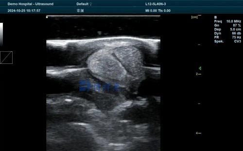
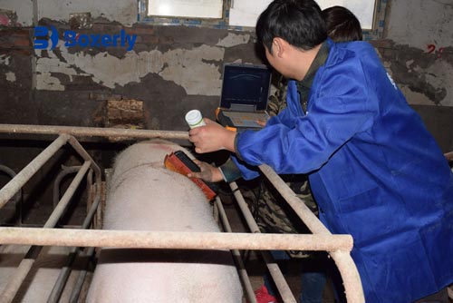
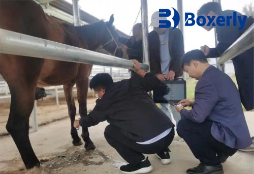
Role of Ultrasonography in Diagnosis
While palpation and visual inspection are helpful, ultrasonography is the gold standard for diagnosing tendon injuries in horses. The real-time image allows vets to see:
-
The exact location and size of lesions
-
The degree of fiber disruption
-
The stage of healing over time
A skilled vet will scan the tendon in both transverse and longitudinal planes, comparing the injured leg with the sound one. This not only confirms the diagnosis but also provides a baseline for tracking progress.
Here’s a simplified example of what vets typically assess on an ultrasound scan:
| Parameter | What It Shows | Why It Matters |
|---|---|---|
| Cross-sectional area (CSA) | Size of the tendon at injury site | Indicates swelling or tissue loss |
| Echogenicity | Brightness of tendon fibers | Hypoechoic (dark) areas suggest fiber damage |
| Fiber alignment | Pattern of collagen fibers | Poor alignment means slower healing |
| Vascularity (Doppler) | Blood flow in healing tissue | Higher flow indicates active repair |
Treatment and Management Strategies
Once the diagnosis is confirmed, management is usually a blend of rest, controlled exercise, and targeted therapies.
1. Acute Phase (First 7–10 Days)
-
Strict stall rest
-
Cold therapy 2–3 times daily to reduce inflammation
-
Support bandaging to limit swelling
-
Non-steroidal anti-inflammatory drugs (NSAIDs) as prescribed
2. Sub-Acute and Healing Phase
-
Gradual introduction of hand-walking
-
Regular ultrasound rechecks every 4–6 weeks
-
Controlled turnout only when safe
-
Therapeutic shoeing to reduce tendon strain
3. Regenerative Therapies (Optional but Increasingly Popular)
-
Platelet-Rich Plasma (PRP) injections
-
Stem cell therapy
-
Shockwave therapy for stimulating healing
These treatments, widely discussed in European and North American equine circles, can help improve fiber quality and reduce re-injury risk, though costs vary and results depend on the case.
Rehabilitation: The Long Road Back
One of the hardest parts for owners is sticking to the slow rehab schedule. Tendons heal far more slowly than muscle because of their limited blood supply. Most vets recommend 6–12 months before full return to work.
A typical progression might look like this:
-
First 6–8 weeks – Stall rest with hand-walking
-
Next 2–3 months – Increase walking duration, add short trot sets if ultrasound looks good
-
Final months – Gradual return to canter, then sport-specific training
Rushing this process almost always leads to setbacks.
Prevention: Keeping Tendons Healthy
While not all injuries can be prevented, some habits make a big difference:
-
Build fitness gradually; avoid sudden workload spikes
-
Warm up and cool down properly
-
Keep hooves balanced and well-trimmed
-
Choose consistent, safe footing for training
-
Schedule regular veterinary checks for high-performance horses
In the words of a British eventing vet I once spoke with, “Preventing tendon injuries is about respecting the horse’s biological limits—not just its competitive drive.”
Final Thoughts
Equine tendon injuries can be career-changing events, but with early diagnosis, proper use of ultrasonography, and disciplined rehabilitation, many horses return to a good level of performance. The process demands patience from both horse and handler, but every small milestone—whether it’s a clean ultrasound scan or the first sound trot—makes the journey worth it.