Diagnostic Value of Ultrasound in Detecting Tendon Injuries in Performance Horse Limbs
In the world of equine sports medicine, diagnosing soft tissue injuries with accuracy and speed is critical to the performance and longevity of elite horses. Tendon injuries, particularly in the limbs, are among the most common and debilitating conditions faced by performance horses, including racehorses, show jumpers, and eventers. These injuries can lead to long rehabilitation periods, financial loss, and, in severe cases, career-ending outcomes.

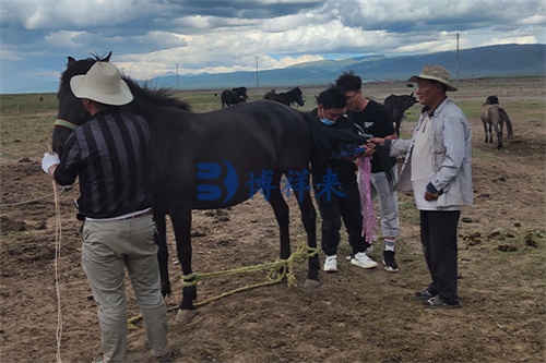
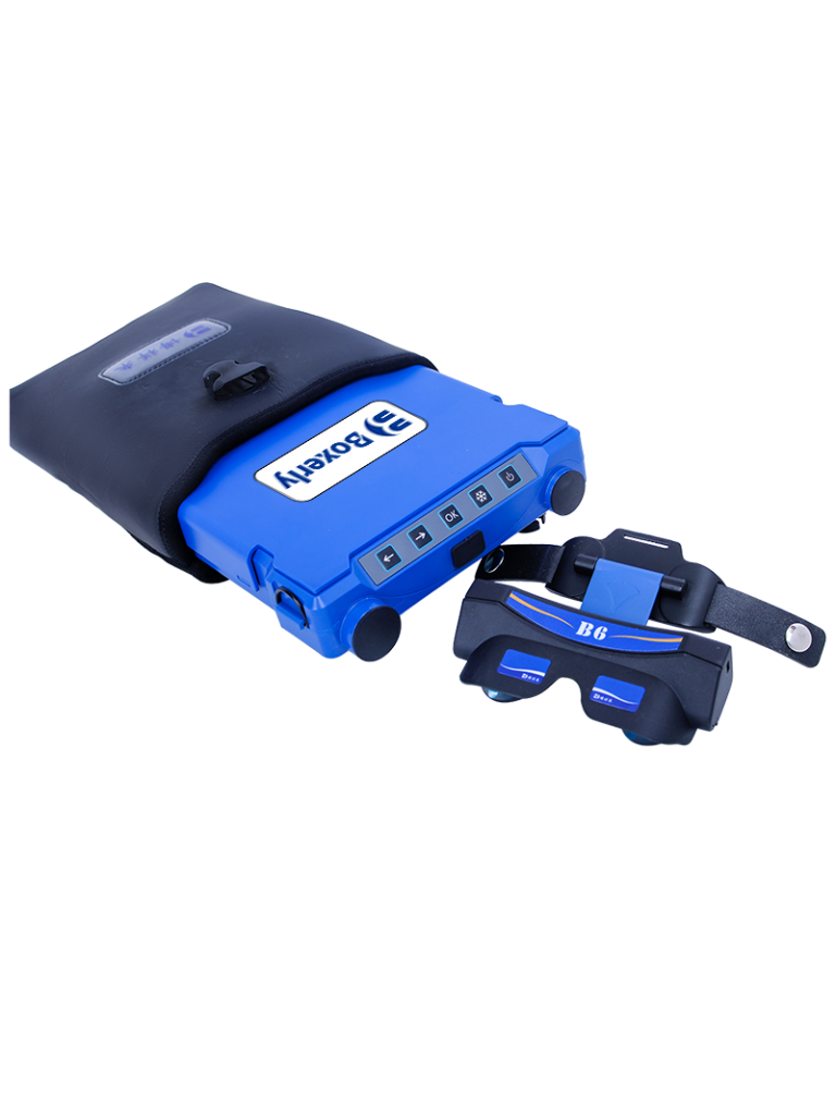
Ultrasound, a non-invasive and highly accessible imaging technique, has become the gold standard for assessing tendon integrity in horses. Thanks to advancements in veterinary imaging, portable devices now enable on-site, real-time diagnostics with exceptional detail. This article explores how equine veterinarians in both research and clinical practice—especially in countries with advanced equine sports medicine such as the United States, the United Kingdom, and Australia—use ultrasound to evaluate, monitor, and manage tendon injuries in equine limbs.
Understanding Tendon Injuries in Performance Horses
Tendons are strong fibrous connective tissues that connect muscles to bones and are crucial in facilitating locomotion. In equine athletes, the superficial digital flexor tendon (SDFT) and deep digital flexor tendon (DDFT) in the limbs are especially prone to injury due to repetitive strain, overextension, and high-speed performance.
Common tendon injuries in performance horses include:
-
Tendinitis (inflammation of the tendon)
-
Tendon rupture or partial tear
-
Tendon sheath inflammation (tenosynovitis)
-
Scar tissue development from previous injury
These injuries often present with subtle signs at first—mild lameness, swelling, or heat in the limb—which can be missed without detailed imaging. This is where ultrasound becomes an invaluable diagnostic tool.
Why Ultrasound is Ideal for Equine Tendon Assessment
Ultrasound offers several distinct advantages when diagnosing tendon injuries in horses:
-
Non-invasive and portable: Unlike MRI or CT, which require expensive, stationary equipment and sedation or anesthesia, ultrasound can be performed stall-side with minimal restraint.
-
Real-time imaging: It allows veterinarians to assess tendon structure, measure lesion size, and observe fiber alignment instantly.
-
Safe and repeatable: Ultrasound does not emit ionizing radiation, making it safe for repeated follow-ups.
-
Cost-effective: Especially in field practice, ultrasound provides detailed diagnostic capabilities at a fraction of the cost of other advanced imaging techniques.
These features have made ultrasound indispensable in equine practices across the globe, particularly in sports medicine and rehabilitation programs.
Ultrasound Imaging: How It Works for Tendon Evaluation
Veterinary ultrasound systems operate primarily using B-mode (brightness mode), which creates two-dimensional grayscale images by interpreting the intensity of sound waves reflected from different tissue types. Tendons appear as parallel, hyperechoic (bright) linear structures in a normal scan.
When a tendon is injured:
-
Hypoechoic (dark) areas may appear, indicating fluid or tissue damage.
-
Disruption of fiber alignment is visible, showing the extent and pattern of the tear.
-
Increased cross-sectional area may suggest swelling or inflammation.
-
Fibrosis or calcification from older injuries may present as hyperechoic spots or irregular structures.
For performance horses, these indicators are critical in evaluating the severity of the injury and determining prognosis.
Diagnostic Protocol in the Field
Ultrasound tendon exams in horses typically follow a structured scanning protocol:
-
Preparation: The limb is clipped and cleaned, and coupling gel is applied for optimal probe contact.
-
Scanning zones: The tendon is imaged in both longitudinal and transverse planes from just below the knee/hock to the pastern, divided into numbered zones (e.g., zones 1A, 1B, etc.) for consistent reporting.
-
Lesion assessment: The clinician measures lesion size (length, width, depth), fiber pattern disruption, and echogenicity.
-
Grading the injury: Some veterinarians use established grading systems (e.g., a 0–3 scale for severity) to classify the injury.
Veterinarians in the UK and Australia have further refined these methods by integrating Elastography—a technique that assesses tendon elasticity—and Power Doppler to evaluate blood flow in healing tissues.
International Perspectives on Tendon Ultrasound Use
United States
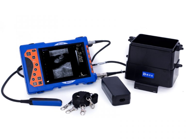
In the U.S., ultrasound is the first-line imaging modality for equine limb injuries, with equine sports medicine clinics routinely performing tendon evaluations. Portable systems like the BXL-V50 or the SonoSite Edge are widely used in racetracks, polo stables, and training barns. Many American practitioners also use ultrasound to guide regenerative treatments, such as PRP (Platelet-Rich Plasma) or stem cell injections, directly into the lesion.
United Kingdom
British veterinarians emphasize structured rehabilitation following diagnosis. The Royal Veterinary College encourages weekly or bi-weekly ultrasound checks during the first three months of tendon injury healing, ensuring progressive and monitored fiber regeneration.
Australia
Given the high prevalence of tendon injuries in Australian Thoroughbreds, ultrasound is considered essential in early detection. Equine hospitals often combine ultrasound with shockwave therapy or laser therapy, timing treatments based on serial scans.
Monitoring Recovery and Return to Work
Ultrasound does not only serve in initial diagnosis but is crucial in long-term monitoring. Healing tendons exhibit gradual changes over time:
-
Restoration of echogenicity
-
Re-alignment of fibers
-
Decrease in lesion size
Veterinarians typically perform serial scans every 4–6 weeks to adjust rehabilitation protocols. A common benchmark is 70% echogenic fiber re-alignment, considered the minimum for initiating trotting under saddle.
This dynamic, image-guided approach significantly reduces the risk of re-injury, which, according to studies, occurs in over 40% of performance horses if the tendon has not healed fully before returning to intense activity.
Limitations and Future Advancements
While ultrasound is highly effective, it does have limitations:
-
Operator dependency: Diagnostic accuracy depends on the clinician’s experience and skill.
-
Limited depth: Deeper structures in large limbs may not be visualized clearly.
-
Resolution vs. portability trade-off: While portable units like the BXL-V50 offer excellent field performance, higher-end cart-based systems can offer greater image detail.
Emerging technologies like 3D ultrasound, AI-driven pattern recognition, and ultrasound elastography are being explored in research settings and may become mainstream in the next few years.
Conclusion
Tendon injuries in performance horses pose significant challenges for veterinarians and owners alike. Early, accurate diagnosis is key to a successful outcome, and ultrasound has proven to be the most reliable, versatile, and practical imaging tool for this purpose.
By offering real-time, repeatable, and non-invasive evaluation of tendon structure and healing progress, ultrasound has revolutionized how we care for athletic horses. International practices, from the U.S. to Australia, demonstrate its critical role not just in diagnosis, but also in guiding treatment and reducing relapse.
As technology advances and more portable yet powerful systems become available, ultrasound will remain the cornerstone of equine tendon diagnostics—ensuring that performance horses have the best chance at recovery and a return to their competitive careers.
References
-
Denoix, J.-M. (2020). Ultrasound Imaging of Tendons in Horses. Equine Veterinary Journal.
-
Dyson, S., & Murray, R. (2019). Diagnosis and Management of Tendon Injuries in the Equine Athlete. The Veterinary Clinics of North America: Equine Practice.
-
Royal Veterinary College. (2022). Ultrasound in Equine Lameness Evaluation. https://www.rvc.ac.uk/research/research-centres-and-facilities/equine
-
American Association of Equine Practitioners (AAEP). (2021). Guidelines on Diagnostic Imaging in Equine Practice.
-
Equine Veterinarians Australia (EVA). (2023). Using Ultrasound to Diagnose and Monitor Tendon Injuries.


