The Critical Role of Veterinary Ultrasound in Diagnosing and Managing Equine Tendon Injuries
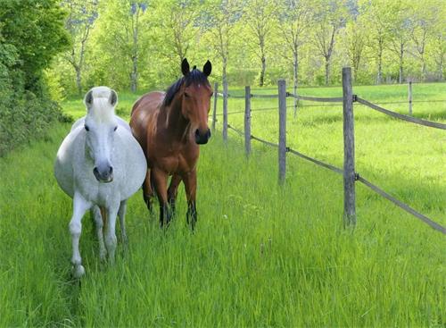
As any equine veterinarian or horse owner knows, tendon health is paramount to a horse’s athletic performance and overall well-being. Tendon injuries are among the most common musculoskeletal problems affecting horses, especially those involved in sport, racing, or heavy work. Early and accurate diagnosis, followed by timely intervention, is key to successful recovery and prevention of long-term complications.
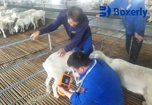
Among the advanced diagnostic tools available today, veterinary ultrasound has become an indispensable technology for evaluating equine tendon structure, detecting injuries, and monitoring healing progress. This non-invasive, real-time imaging technique offers unprecedented insight into tendon fiber integrity, lesion extent, and subtle changes invisible to other examination methods.
In this article, I will explore how veterinary ultrasound is applied in equine tendon assessment, the benefits it offers, and why it is widely recognized by veterinary professionals around the world as the gold standard for tendon diagnostics and management.
Understanding Equine Tendon Anatomy and Common Injuries
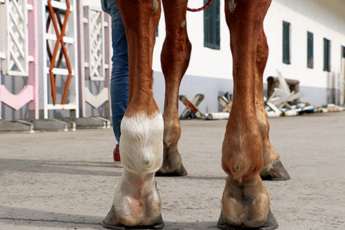
Tendons are dense bands of connective tissue that connect muscle to bone, transmitting the force required for movement. In horses, the superficial digital flexor tendon (SDFT) and deep digital flexor tendon (DDFT) in the limbs are particularly susceptible to injury due to repetitive strain and impact.
Tendon injuries in horses range from mild inflammation (tendinitis) to partial tears and complete ruptures. Common causes include overexertion, improper shoeing, trauma, or conformation defects. Left untreated or poorly managed, tendon injuries can lead to chronic lameness or career-ending damage.
Traditional clinical examination methods—palpation, gait observation, and thermography—can suggest tendon problems but lack the precision to define injury extent or guide prognosis reliably. This is where veterinary ultrasound imaging revolutionizes diagnosis.
The Advantages of Veterinary Ultrasound in Tendon Evaluation
Ultrasound provides a window into the internal structure of tendons without surgery or radiation. It works by emitting high-frequency sound waves that reflect off tissues, creating detailed images of tendon fibers, lesions, and surrounding structures.
Some key advantages recognized by veterinarians globally include:
-
Non-invasive and safe: Ultrasound scanning is painless, poses no radiation risk, and can be repeated frequently to monitor healing.
-
High-resolution imaging: Modern ultrasound devices offer high-definition images that reveal fiber alignment, core lesions, and new blood vessel formation associated with healing.
-
Real-time dynamic assessment: Tendons can be evaluated during movement or flexion, allowing assessment of functional integrity.
-
Quantitative evaluation: Measuring lesion size, echogenicity (brightness), and fiber disruption enables objective monitoring of injury progression or repair.
-
Early detection: Ultrasound can identify subtle tendon fiber fraying or thickening before clinical signs worsen.
How Ultrasound Examination of Equine Tendons is Performed
A typical ultrasound exam for tendons involves:
-
Preparation: The limb is cleaned and shaved to ensure proper probe contact.
-
Positioning: The horse is usually sedated and placed in a comfortable standing position.
-
Probe selection: A high-frequency linear transducer (typically 7.5–12 MHz) is used for detailed tendon imaging.
-
Scanning planes: Tendons are scanned longitudinally and transversely at multiple points along their length.
-
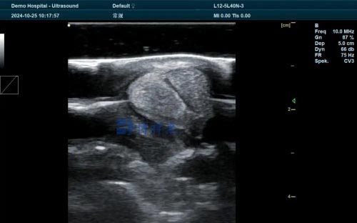
Image interpretation: Healthy tendons appear as uniform, tightly packed parallel fibers with medium echogenicity. Lesions disrupt this pattern, appearing hypoechoic (dark areas) or anechoic (black fluid-filled zones).
Interpreting Ultrasound Findings in Tendon Injuries
Veterinary professionals classify tendon injuries on ultrasound by:
-
Lesion size: Cross-sectional area affected, often expressed as a percentage of the total tendon area.
-
Lesion echogenicity: Hypoechoic regions indicate fiber tearing or inflammation; hyperechoic areas can indicate fibrosis or calcification.
-
Fiber alignment: Loss of normal parallel fiber pattern correlates with severity.
-
Neovascularization: New blood vessel growth is a marker of healing but can also indicate chronic inflammation.
This detailed characterization helps veterinarians tailor rehabilitation programs, including controlled exercise, shockwave therapy, or platelet-rich plasma injections.
Monitoring Tendon Healing with Ultrasound
One of the most valuable aspects of ultrasound is tracking tendon recovery over time. Regular scanning sessions provide objective data on lesion resolution, fiber realignment, and scar tissue formation.
Veterinarians and trainers can then adjust rehabilitation protocols to optimize healing without risking re-injury. In fact, studies from equine veterinary research institutions worldwide confirm that ultrasound-guided rehab improves long-term outcomes compared to clinical evaluation alone.
The Impact of Veterinary Ultrasound on Equine Sports Medicine
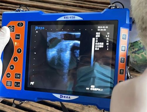
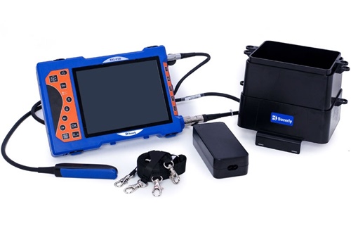
The widespread adoption of ultrasound in equine sports medicine reflects its effectiveness in both diagnosis and prognosis. Leading equine hospitals and research centers use portable ultrasound units, such as the BXL-V50, which combine high image quality with field durability, allowing on-site examinations at training yards and competitions.
This portability means veterinarians can promptly evaluate tendon status after an injury incident, make immediate management decisions, and provide owners with clear, evidence-based prognoses.
Comparing Ultrasound with Other Imaging Modalities
While MRI and thermography also contribute to tendon assessment, ultrasound offers distinct advantages:
-
Cost-effectiveness: Ultrasound devices are generally more affordable and accessible.
-
Ease of use: Portable units can be operated in the field by trained veterinarians.
-
Dynamic capability: Unlike MRI, ultrasound can image tendons during movement.
-
No sedation requirement: Many ultrasound exams can be performed with minimal sedation.
Thus, ultrasound remains the frontline imaging tool for tendon evaluation globally.
Limitations and Challenges of Ultrasound in Tendon Imaging
Despite its many benefits, ultrasound does have limitations:
-
Operator skill greatly affects image quality and interpretation accuracy.
-
Deep or obscured tendons may be difficult to visualize fully.
-
Early microscopic fiber damage might be undetectable.
-
Differentiating scar tissue from healthy tendon requires experience.
Ongoing advances in ultrasound technology, such as Doppler imaging to assess blood flow and elastography to evaluate tissue stiffness, are helping to overcome these challenges.
International Perspectives and Research on Equine Tendon Ultrasound
Studies from veterinary schools in Europe, North America, and Australia consistently highlight ultrasound as essential in equine tendon management. Research published in journals like the Equine Veterinary Journal and Veterinary Radiology & Ultrasound underlines:
-
The predictive value of lesion size and echogenicity on return-to-performance.
-
The benefit of ultrasound-guided therapies.
-
The role of ultrasound in injury prevention programs through pre-season screening.
This broad scientific consensus reinforces the global veterinary community’s reliance on ultrasound for tendon care.
Practical Tips for Veterinarians and Horse Owners
To maximize ultrasound’s benefits in tendon health:
-
Ensure examinations are done by skilled ultrasonographers familiar with equine anatomy.
-
Combine ultrasound findings with clinical examination and history.
-
Schedule serial ultrasounds during rehabilitation for monitoring.
-
Use ultrasound data to guide controlled exercise progression and therapy choice.
-
Educate owners on ultrasound findings for informed care decisions.
Conclusion
In the world of equine health, maintaining tendon integrity is crucial to a horse’s performance and longevity. Veterinary ultrasound has revolutionized how tendon injuries are diagnosed, treated, and monitored. Its non-invasive nature, detailed imaging capability, and real-time assessment make it an indispensable tool in modern equine practice.
From early injury detection to guiding rehabilitation and predicting outcomes, ultrasound empowers veterinarians to deliver targeted, effective care. As portable ultrasound technology like the BXL-V50 becomes increasingly accessible, its role in safeguarding equine musculoskeletal health will only grow stronger.
For horse owners, trainers, and vets alike, embracing veterinary ultrasound in tendon examination represents a leap forward toward healthier, more resilient horses.
References
-
Smith, R. K. W., & McIlwraith, C. W. (2014). Diagnosis and Management of Tendon and Ligament Injuries in Horses. Equine Veterinary Journal, 46(6), 689–697. https://doi.org/10.1111/evj.12267
-
Dyson, S. J. (2015). Ultrasonography in Equine Tendon Injuries: Current Perspectives. Veterinary Radiology & Ultrasound, 56(2), 125-133. https://doi.org/10.1111/vru.12202
-
O’Grady, S. E., & Williams, J. M. (2018). Clinical Use of Portable Ultrasound for Tendon Injury Assessment in Horses. Journal of Equine Veterinary Science, 67, 62-69. https://doi.org/10.1016/j.jevs.2018.06.011
-
Equine Veterinary Education. (2022). Latest Advances in Equine Tendon Ultrasound Imaging. https://www.equinedveterinaryeducation.com/articles/latest-tendon-ultrasound-advances