Ultrasound Imaging Characteristics During Sow Pregnancy
Understanding reproductive progress in sows has become a pivotal part of modern pig production. As gestation monitoring technology advances, ultrasound imaging—especially B-mode ultrasound—has become an indispensable tool for confirming pregnancy, detecting reproductive issues, and ensuring healthy fetal development. Unlike invasive methods, ultrasound offers a safe, repeatable, and accurate method of evaluating sow pregnancy progression. While the underlying technology is widely adopted in veterinary practice worldwide, interpreting early-stage pregnancy images in pigs requires years of hands-on experience and practical pattern recognition.

In this article, we explore the sonographic characteristics of sow pregnancy from the earliest detectable signs to more advanced gestation stages. We also compare ultrasound images of normal pregnancy to those of uterine pathologies such as pyometra and discuss common diagnostic pitfalls faced by both new and experienced practitioners.
The Early Use of Ultrasound in Sow Reproduction
Veterinary practitioners in developed pig-producing countries such as Denmark, the Netherlands, the United States, and Canada have incorporated ultrasound technology into daily breeding routines for decades. Ultrasound machines are commonly used to detect pregnancy, confirm embryo viability, and monitor uterine health. However, many swine specialists emphasize that understanding sow ultrasound images, especially in the first three weeks after insemination, is more of an art than a science.
Around day 18 post-mating, an experienced operator may detect faint echogenic signals on the monitor—often described by veterinarians as “grain-like shadows on a glass surface”. This faint speckling represents early embryonic fluid collection and uterine activity. At day 19, multiple black, round fluid-filled structures the size of mung beans may be observed. These are early embryonic vesicles.
By day 20, sow pregnancy images become more distinct. Dark, round areas about the size of soybeans appear, and their edges are more clearly defined. Still, distinguishing these from abnormal uterine contents such as pus requires caution and experience.
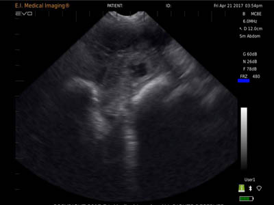
Recognizing Patterns by Gestational Day
Day 21
The image clarity improves, with larger and more rounded anechoic (black) areas, indicating growing embryonic sacs. These are often seen in clusters and spaced evenly throughout the uterine horns.
Day 24
The black fluid areas resemble small coins in size, and the edges are more defined, making diagnosis more straightforward. From this stage onward, most trained ultrasound users can reliably confirm pregnancy.
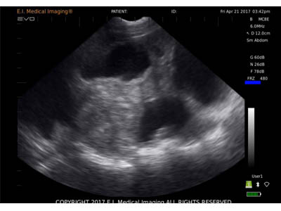
Day 30
At this stage, the round black embryonic sacs are significantly enlarged. Not only are they clear and well-circumscribed, but their growth pattern also becomes easier to differentiate from any pathological uterine fluid collection. The embryo may begin to be visible within the sac.
Day 35 onward
With increasing gestation, the embryonic shape becomes visible, and the fetus begins to resemble a piglet in formation. Head, spine, and even limb buds might appear as white echoes within the dark fluid, confirming fetal health and development.
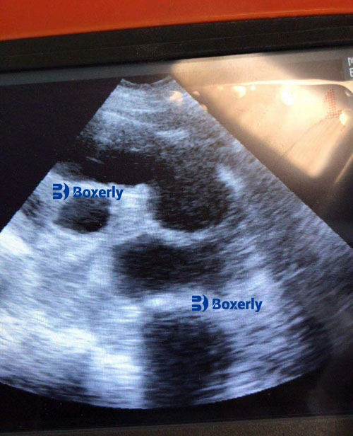
Differentiating Normal Pregnancy from Uterine Disorders
Uterine Pyometra vs. Early Pregnancy (Day 18–20)
Pyometra (uterine pus accumulation) is one of the main conditions that can mimic early pregnancy ultrasound features. On-screen, pyometra may appear as several uneven, low-echo or anechoic areas. However, unlike embryonic sacs, the edges are poorly defined, and the size distribution is inconsistent. Fluid content may also vary in density. These features help practitioners make a more confident diagnosis.
Pregnancy vs. Full Bladder Post-Day 30
Beyond 30 days of gestation, the full urinary bladder can present a single large, black oval structure—similar to an embryonic sac. However, the key differentiator is quantity: a pregnant uterus presents multiple black circular zones, whereas the bladder appears as a single anechoic mass. Additionally, if the bladder contains excessive urine, it may obscure the uterine field altogether, requiring the sow to void before a clear image can be captured.
Practical Field Experience and Training
In North America and Europe, many pig farms invest in hands-on ultrasound training programs. According to a 2023 report by the European Association of Swine Practitioners (EASP), nearly 75% of commercial pig farms use portable ultrasound scanners during early pregnancy checks. However, only about 40% of farm technicians report full confidence in interpreting images before day 22.
Dr. Heather Monaghan, a swine reproduction specialist in Ontario, Canada, emphasizes the importance of building a personal image database. “Each sow, scanner, and scanning angle is slightly different. Documenting what you see at each stage helps build recognition patterns faster,” she notes.
In Asian pig farms, particularly in China and Vietnam, ultrasound is increasingly integrated into AI (Artificial Insemination) programs. Local technicians often share video logs of real-time scans and create comparison libraries of pregnant vs. non-pregnant sows to assist newcomers in shortening their learning curve.
Advantages of Using Ultrasound in Sow Management
-
Non-Invasive & Stress-Free
Ultrasound doesn’t harm the animal or interfere with pregnancy progression. Sows remain calm, and repeated scanning is safe. -
Early Pregnancy Confirmation
As early as day 18, skilled users can detect changes, improving farm productivity by quickly rebreeding non-pregnant animals. -
Health Monitoring
Beyond pregnancy, ultrasound is used to check for uterine infections, cysts, and abnormal fluid collections, helping maintain herd reproductive health. -
Economic Benefit
Detecting pregnancy earlier saves feed costs and reduces non-productive sow days (NPD), which is one of the key performance indicators on commercial pig farms.
Recommendations for Beginners
-
Start scanning at day 24 if you are new to ultrasound. The images are more distinct and harder to confuse with other structures.
-
Scan in a low-light environment, which improves image visibility on the ultrasound monitor.
-
Use a generous amount of ultrasound gel to ensure proper probe-skin contact and avoid image dropout.
-
Keep notes and save images for future comparison. Over time, you will notice subtle differences that become intuitive.
Conclusion
Ultrasound imaging has revolutionized how we manage sow reproduction. From detecting pregnancy at the earliest stage to differentiating between normal and pathological conditions, the value of this tool lies in its real-time insight, safety, and cost-saving potential. However, as with many diagnostic methods, successful interpretation requires not just access to technology but also continuous experience and observation.
As more swine farms across the world embrace precision farming techniques, ultrasound will remain a cornerstone technology—not just for pregnancy checks, but for reproductive health monitoring and herd-level decision-making. In time, even beginner users will gain confidence by understanding the evolving image patterns throughout the sow’s pregnancy, ultimately boosting productivity, animal welfare, and farm profitability.
Reference Sources:
-
Monaghan, H. (2023). “Identifying Early Pregnancy and Uterine Health in Sows Using B-Mode Ultrasound.” Canadian Journal of Swine Veterinary Practice, 45(2), 117–123.
https://www.cjsvp.org/early-pregnancy-sow-ultrasound -
European Association of Swine Practitioners (2023). “Ultrasound Use in Commercial Pig Farms: A Continental Survey.”
https://www.easvp.org/resources/ultrasound-in-pig-farms -
National Hog Farmer (2022). “Improving Reproductive Efficiency in Sows with Ultrasound Technology.”
https://www.nationalhogfarmer.com/reproduction/ultrasound-reproductive-efficiency