Portable Ultrasound Enhancing Disease Diagnosis Efficiency at Farms
Have you ever thought about how a small, handheld device could transform animal health management on your farm? Gone are the days when disease detection meant waiting for visible symptoms or relying solely on blood tests. Today, portable ultrasound—think of devices like the BXL‑V50—brings real-time insights right to the barn aisle. Let’s dive into how these gadgets are reshaping the way farmers and vets team up to keep livestock healthy, performing under peer‑to‑peer chat vibes rather than stiff textbook style.

Why Early Detection Matters
Timely intervention makes all the difference when an animal falls ill. Delays in spotting conditions like mastitis, pneumonia, or joint infections can lead to serious welfare concerns and hefty treatment costs. Imagine catching a mild lung lesion or early‑stage uterine infection before antibiotics become less effective. With portable ultrasound, you’re essentially wielding a window into tissues and organs, spotting problems before they spiral out of control. It’s like having a mini diagnostic lab strapped to your hip—only you don’t need a lab coat.
What Makes Portable Ultrasound Tick
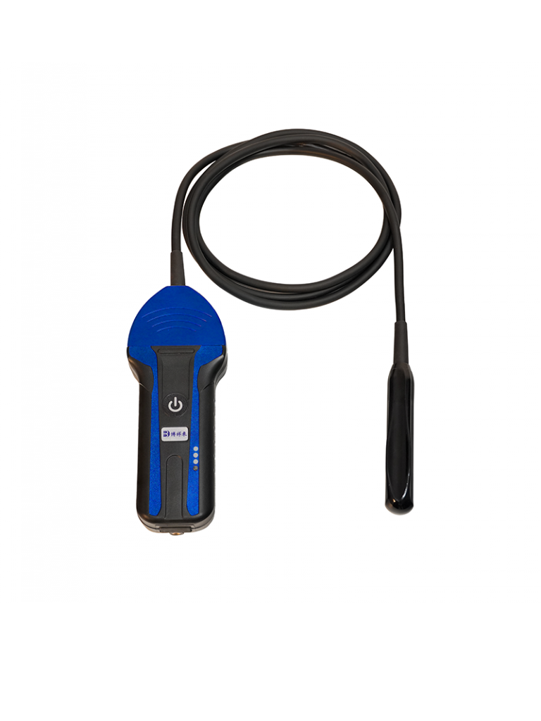
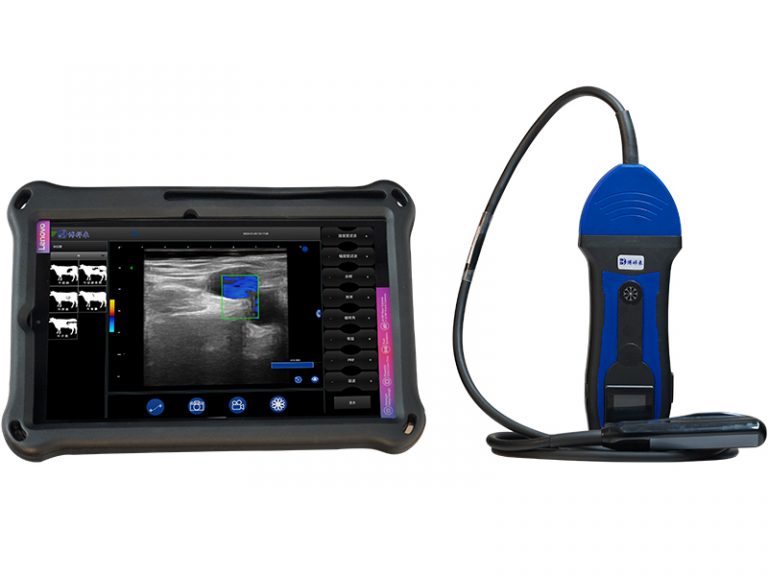
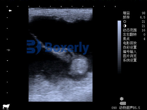
At its core, ultrasonography sends high‑frequency sound waves into the animal’s body and captures echoes as tissues reflect them back. Those echoes translate into on‑screen images of muscle layers, organ surfaces, fluid pockets, and more. Unlike stationary machines, portable units combine compact probes, rechargeable batteries, and intuitive software to deliver B‑mode scans wherever you need them—be it in the field, at the milking station, or even in remote grazing areas.
-
Portability: Weighing under two pounds, devices like the BXL‑V50 slip easily into a jacket pocket.
-
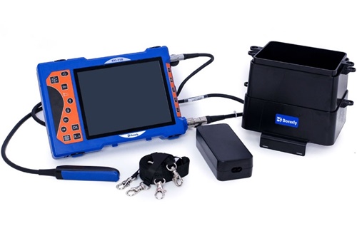
Durability: Rugged casings and IP‑rated seals shrug off dust, water splashes, and occasional barnyard bumps.
-
Battery Life: With up to seven hours on a single charge, you won’t cut your day short hunting for outlets.
-
User Interface: Touchscreen controls and preset animal‑specific settings get you scanning fast, even if you’re not an imaging expert.
Spotting Mastitis Before It Hits Hard
Mastitis remains a top headache for dairy producers. Traditional checks—udder palpation, milk somatic cell counts—often lag behind the actual infection timeline. Portable ultrasound steps in to visualize early tissue edema, abscess formation, and ductal changes. On my farm, scanning each quarter twice a week helped me flag two subclinical cases before any drop in milk yield. Treatment kick‑off became quicker, antibiotic use more targeted, and overall somatic cell counts plummeted.
-
Prepare the udder: Clean and apply coupling gel.
-
Probe placement: Position the linear array probe at the gland‑front junction.
-
Interpretation: Hypoechoic (dark) streaks signal tissue fluid; hyperechoic (bright) spots can hint at fibrosis or abscess.
-
Action: Early antibiotic therapy or selective dry cow treatment based on scan findings.
Lung Health: Beyond the Stethoscope
Auscultation only scratches the surface when it comes to respiratory issues. Portable ultrasound lets you scan intercostal spaces, looking for pleural line irregularities, comet‑tail artifacts, or consolidation zones that mark pneumonia. In young calves, where respiratory disease can escalate rapidly, this capability is a game‑changer. You can differentiate viral from bacterial patterns, choose appropriate therapies, and monitor recovery—all without moving the calf to a clinic.
-
Normal lung: Sliding pleura and reverberation artifacts.
-
Early pneumonia: Small fluid pockets lining the pleura.
-
Advanced consolidation: Tissue‑like echotexture replacing air‑filled alveoli.
Joints, Tendons, and Beyond
Lameness costs the industry billions annually, yet many cases start as subtle tendon or ligament strains. With a portable ultrasound unit, examining fetlock, hock, or stifle tendons becomes straightforward. A quick scan can reveal tendon fiber disruptions, sheath effusions, or early osteoarthritis changes. Armed with this intel, you can tweak hoof trims, adjust exercise regimens, or initiate anti‑inflam treatments well before visible lameness sets in.
Reproductive Health on the Fly
Whether you’re synchronizing heifers or diagnosing repeat breeders, real‑time feedback matters. Transrectal ultrasonography provides clear images of ovarian follicles, corpora lutea, and uterine horns. You can confirm pregnancies as early as 28 days post‑AI, differentiate embryonic loss from pseudopregnancy, and spot uterine infections that would otherwise go unnoticed. By integrating these scans into your breeding protocol, you optimize calving intervals and enhance herd fertility metrics.
Bringing It All Together: Workflow Integration
Let’s keep it practical. Portable scanners earn their keep when woven into daily routines:
-
Morning health checks: A five‑minute scan during feeding assessments.
-
Record keeping: Capture and store images via integrated USB or wireless upload.
-
Team collaboration: Share scans with your vet in real time for remote consultations.
-
Continuous training: Use on‑device tutorials or quick‑reference guides to sharpen technique.
One farm I know set up weekly “scan‑n‑share” sessions, where staff review interesting cases together—everything from early metritis to mild tendonitis. This peer‑learning approach not only raises everyone’s skills but also fosters a culture of proactive health management.
Overcoming Common Challenges
Certainly, there’s a learning curve. Interpreting echoes and differentiating normal from pathological patterns takes practice. But most portable ultrasound makers offer online training modules, animal‑specific presets, and manufacturer support hotlines. Also, don’t hesitate to team up with your local vet school or imaging specialist for a weekend workshop. A few hands‑on hours can cut weeks off your mastery timeline.
Where We’re Headed: AI and Automation
Here’s a peek into the future: AI‑powered image analysis is steadily making its way onto portable units. Imagine scanning a udder quarter and having the device highlight suspicious areas or even estimate somatic cell count. Early trials show promise in automated pneumonia detection and tendon lesion grading. As processing power grows, these smart features will transform portable ultrasound from a mere imaging tool into a decision‑support powerhouse.
Wrapping Up
It’s clear that portable ultrasound isn’t just another gadget—it’s a practical bridge between observation and action. By slashing diagnostic delays, sharpening treatment choices, and boosting overall herd health, these devices deliver real ROI. Whether you’re milking dairy cows at dawn or checking piglets in the afternoon, a handheld ultrasound scanner like the BXL‑V50 can be your most trusted ally in disease management.
References
-
Braun, U., & Marmier, O. (2020). Ultrasonography of the Bovine Mammary Gland. Veterinary Radiology & Ultrasound. https://onlinelibrary.wiley.com/doi/10.1111/vru.12811
-
Ollivett, T. L., & Buczinski, S. (2016). On-farm use of ultrasonography for bovine respiratory disease diagnosis. Veterinary Clinics of North America: Food Animal Practice. https://www.sciencedirect.com/science/article/pii/S074907201630022X
-
Laven, R. A., & Peters, A. R. (2000). Reproductive ultrasonography in cattle. Animal Reproduction Science. https://www.sciencedirect.com/science/article/pii/S0378432000001808