
Why Is Ultrasound Transmitted in Pulses?
Ultrasound has become one of the most valuable diagnostic tools in veterinary medicine, particularly for large animals. Its ability to provide real-time, non-invasive, and highly detailed images of soft tissue structures makes it indispensable in both clinical and farm environments. A frequently asked question among livestock owners and veterinary students is: Why is ultrasound transmitted in pulses instead of a continuous wave? As a livestock farmer who regularly relies on ultrasound examinations to monitor herd health and reproduction, I can confidently say—yes, ultrasound is transmitted in pulses, and there are solid scientific and practical reasons behind this design.
Understanding Pulsed Ultrasound Transmission
An ultrasound image is formed by sending wave pulses in different directions. When a pulse travels through the body and encounters a structure with a different acoustic density—such as a cell, a group of cells, or denser tissues like tumors or organs—it reflects back toward the probe. The transducer, which initially emitted the pulse, then switches to receiving mode and picks up the returning echoes.
The key to building a useful image lies in determining two critical parameters: direction and distance. Since the system knows the direction in which the pulse was originally sent, it also knows where to expect the echo from. The distance of the reflecting structure is calculated based on the time it takes for the pulse to travel to the object and bounce back. This is known as round-trip travel time. Simply put, the further away a structure is, the longer it takes for the echo to return. By combining direction and distance, the scanner plots a bright spot on the image to represent the location of that structure.
This process would be impossible with continuous wave (CW) ultrasound. In CW systems, the probe continuously emits and receives sound waves without interruption. This makes it very difficult to measure timing, and therefore depth, accurately. That’s why pulsed wave (PW) ultrasound is preferred for imaging, as it allows the machine to “ping” the tissue and “listen” for the echo, calculating exactly where the reflection came from.
Why Pulses Are Necessary in Veterinary Imaging
To uniquely determine the depth of reflectors (structures inside the body), the spacing between the pulses must be long enough to ensure that the returning echoes from one pulse are received before the next pulse is sent. If pulses were emitted continuously or too rapidly, the echoes would overlap, leading to confusing signals and blurred images. This becomes especially important in animals with large or complex body structures, such as cattle, horses, or pigs.
This is one reason why veterinary ultrasound systems rely so heavily on precisely timed pulsed waves—they help create clean, layer-by-layer images of soft tissues, organs, reproductive systems, and even muscles or tendons. In fact, the entire concept is quite similar to how radar works in air traffic control: a pulse is emitted, and the reflected signal is used to determine the position of an object.
How the Technology Works in Practice
When I perform an ultrasound check on a pregnant cow, for example, I first apply transmission gel to ensure proper acoustic contact. The transducer then sends a short pulse of high-frequency sound into the abdomen. As the sound beam passes through tissues of varying densities, some of it is reflected back to the probe. These returning echoes vary in intensity depending on how different the tissues are. A strong echo occurs when the sound crosses from soft tissue to a denser material like bone or fibrous tissue. Weaker echoes may indicate fluid or soft organ tissue.
These echoes are then converted into electrical signals by the machine, which assembles them into a real-time image. The strength of each echo, its time of return, and the direction it came from are all used to map the internal anatomy on the screen. It’s truly fascinating how something so sophisticated becomes a routine tool on the farm.
Advantages of Pulsed Ultrasound for Livestock
-
Depth Accuracy: Pulses allow precise measurement of depth, which is essential for examining deep structures in large animals.
-
Image Clarity: By receiving echoes from only one pulse at a time, the system avoids echo overlap and produces sharper images.
-
Tissue Differentiation: Pulsed transmission provides detailed contrast between different tissues, helping to distinguish between normal and abnormal areas.
-
Safety: Pulsed waves reduce the risk of tissue heating, making them safer for prolonged or repeated use.
In practice, I use ultrasound for many purposes: checking for pregnancy, identifying ovarian structures, evaluating muscle or tendon injuries, and even guiding biopsies. The ability to see inside the animal without surgery improves outcomes and reduces stress for both the animal and the handler.
Common Formats in Veterinary Ultrasonography
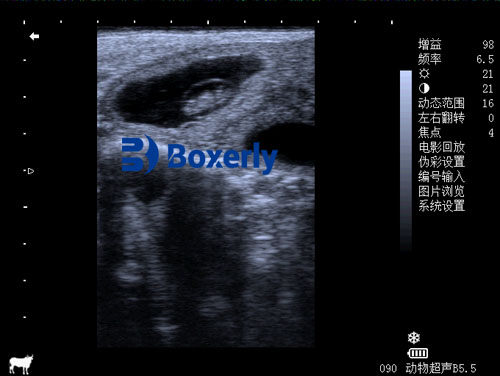
The most commonly used image format is B-mode grayscale. This format creates a two-dimensional image where brightness corresponds to the intensity of the echo. Other formats include M-mode, used for heart scans (echocardiography), and Doppler modes, which evaluate blood flow.
For large animals like cows or horses, we often use low-frequency transducers. These can penetrate deeper into the body but may sacrifice some resolution. For smaller animals or shallow structures, high-frequency probes are used for greater detail.
It’s worth noting that the maximum effective scanning depth is usually around 30 cm. Beyond this, the echoes may be too weak or noisy to interpret. However, when scanning through fluids like urine in the bladder, sound waves travel more efficiently, allowing deeper imaging.
Real-World Application: Reproductive Monitoring in Cattle
On our farm, ultrasound has revolutionized how we manage reproduction. With transrectal ultrasound, we can detect pregnancy as early as 25–30 days post-breeding. We can also monitor the development of follicles and diagnose uterine infections, cysts, or other reproductive issues quickly and accurately. This not only improves herd fertility rates but also saves time and reduces unnecessary treatments.
Summary: Pulses Are the Key to Precision
In conclusion, ultrasound must be transmitted in pulses to allow precise control over timing and image formation. This method enables the machine to map the internal anatomy accurately by calculating both the distance and direction of the reflected echoes. Without pulses, ultrasound would not be able to provide the detailed, real-time diagnostic images that are essential in modern veterinary practice.
As a livestock farmer, I have witnessed firsthand how pulsed ultrasound technology enhances our ability to diagnose, monitor, and treat our animals more effectively. Whether it’s checking on a mare’s pregnancy, evaluating a calf’s heart, or monitoring an old bull’s tendon injury, the benefits of ultrasound are undeniable—and it all begins with a simple, powerful pulse.


Can you be more specific about the content of your article?