Ultrasound Signs Every Horse Owner Should Watch at Farms
For anyone raising horses, whether on a small family farm or a large-scale equine operation, staying one step ahead of health and performance issues can make all the difference. Horses can’t tell us when something’s wrong, which is why tools like ultrasound have become absolute game-changers in recent years. From monitoring pregnancies to catching subtle signs of lameness or internal discomfort, ultrasound allows us to “see” what would otherwise go unnoticed—often before any physical symptoms show up.
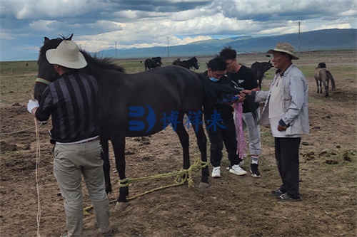
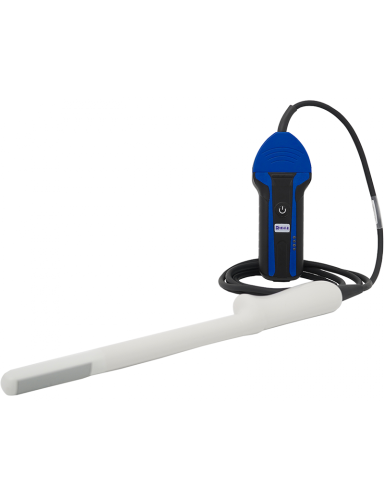
Veterinary ultrasound is no longer limited to clinics. Portable systems are now farm-friendly, rugged, and designed for on-the-go diagnostics. But knowing how to spot the signs worth scanning is just as important as having the machine itself. So let’s walk through the key ultrasound signs every horse owner should be paying attention to—and how they could save you time, money, and heartache.
Watching for Early Pregnancy Clues
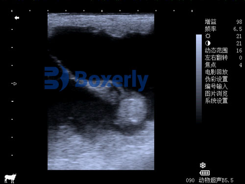
One of the most common uses of ultrasound on horse farms is pregnancy detection. The best time to confirm a successful breeding is usually between 14 and 16 days post-ovulation, when the embryo is visible as a small, round fluid-filled vesicle in the uterus.
Why so early? It’s all about managing twin pregnancies. Mares can occasionally conceive twins, which is risky and often fatal to both embryos. Early ultrasound checks allow for “manual reduction” of one embryo to give the other a better chance at survival. After the initial check, follow-up scans at 25, 35, and 55 days help monitor fetal heartbeat, development, and ensure the pregnancy is progressing normally.
👉 What to watch for:
-
A clear, round black vesicle in the uterus (early pregnancy)
-
Detection of a heartbeat around day 25
-
Uniform uterine tone and absence of fluid pockets (which may signal infection or inflammation)
Uterine Fluid: Not Always Harmless
It’s easy to assume that a little post-breeding uterine fluid is normal—and sometimes it is. But persistent or excessive fluid is one of the main signs of endometritis (uterine inflammation) or infection. On ultrasound, fluid appears black and may range from a thin layer to large accumulations.
Older mares or those with poor uterine clearance are more prone to this issue, and ignoring it can lead to early pregnancy loss or failure to conceive altogether.
👉 Ultrasound signs that should raise concern:
-
Fluid that’s still visible 48+ hours after breeding
-
Fluid pockets with echogenic (grainy) material—often pus or cellular debris
-
Thickened uterine walls indicating inflammation
Regular ultrasound checks during breeding season can help you catch and treat this early, saving the breeding cycle and avoiding expensive delays.
Ovulation Timing and Follicle Monitoring
For successful breeding, timing is everything. Ultrasound gives owners and vets a front-row seat to a mare’s estrous cycle. Follicles in the ovary grow predictably and can be monitored to determine the best time to inseminate. A mature follicle typically measures 35 to 50 mm, and once it reaches this size, ovulation usually occurs within 24–48 hours.
👉 Key signs worth tracking:
-
Rapid follicle growth in the lead-up to ovulation
-
Softening and irregular margins of the follicle (sign it’s about to release the egg)
-
Disappearance of the follicle (meaning ovulation occurred)
-
Corpus luteum formation (evidence that ovulation has happened)
Getting this timing right is especially critical for artificial insemination (AI), where precise hormone management is key to success.
Detecting Lameness and Soft Tissue Injuries
Lameness is one of the costliest and most common issues horse owners face. While hoof testers, flexion tests, and gait analysis are useful, ultrasound offers a direct look at tendons and ligaments that could be silently fraying or strained.
The superficial digital flexor tendon, deep digital flexor tendon, and suspensory ligament are common problem areas. Injuries here often start as microscopic fiber tears and can worsen without visible swelling or heat. With regular ultrasound checks, especially for performance horses, early changes can be spotted before they lead to a full-blown breakdown.
👉 What you’re looking for:
-
Hypoechoic (darker) regions indicating fluid or fiber disruption
-
Enlargement or swelling of tendon structures
-
Irregular fiber pattern suggesting degeneration or scarring
Early rest and rehab guided by ultrasound findings can often save a horse’s career.
Monitoring Foal Development
Once pregnancy is established, there’s still a lot to keep track of. Ultrasound helps evaluate fetal positioning, size, heartbeat, and activity. Between 60 and 120 days, the fetus becomes more active and detailed scans can even pick up anatomical structures like limbs and eyes.
Later on, between 150–240 days, ultrasound is used to measure the thickness of the combined utero-placental unit (CUPT). This helps identify placentitis—a dangerous condition that can lead to abortion. Thicker-than-normal CUPT, combined with signs of fluid buildup or fetal distress, are red flags that call for immediate treatment.
👉 Fetal signs to monitor:
-
Regular, strong fetal heartbeat (over 100 bpm early, then 70–90 bpm later)
-
Consistent fetal movement
-
Normal placental thickness (<13 mm in early mid-gestation, increasing gradually)
Monitoring Postpartum Uterine Involution
After foaling, a mare’s uterus should shrink back to normal within 2–3 weeks. Delays or complications during this period can increase the risk of infection or future breeding problems.
Ultrasound checks during the first 7–10 days postpartum can reveal retained membranes, abnormal fluid, or poor uterine tone.
👉 Ultrasound signs worth noting:
-
Retained placental fragments (visible as echogenic material inside uterus)
-
Excessive fluid post 5 days postpartum
-
Poor or slow reduction in uterine size compared to normal postpartum progression
Catching these issues early allows for quick veterinary intervention before things worsen.
Tracking Body Condition and Muscle Tone
Although ultrasound is typically associated with internal diagnostics, it’s also being used more frequently to track muscle mass and fat coverage, particularly in performance or aging horses. Body condition scoring (BCS) is still useful, but ultrasound offers a more objective look at what’s under the skin.
Using a linear probe, owners or vets can measure longissimus dorsi muscle thickness and subcutaneous fat depth. This can help adjust feeding programs, evaluate fitness, or spot early signs of weight loss before it’s visible.
👉 Signs to monitor:
-
Decreased muscle mass in older or recovering horses
-
Asymmetrical muscle development due to injury or poor saddle fit
-
Changes in fat depth that correlate with energy balance or metabolic conditions
Final Thoughts from the Barn
Ultrasound has become an indispensable tool on many horse farms—not just for reproductive work but for everything from injury monitoring to long-term conditioning. The key is knowing when to scan and what to look for. Whether it’s confirming a pregnancy, catching a sore tendon early, or keeping an eye on fetal health, ultrasound provides valuable information that often translates into better decisions and healthier horses.
And the best part? It’s safe, stress-free, and can be done right in the stall aisle.