Ultrasound Use in Small Animal Practice: Insights from an Experienced Vet
Ultrasound technology has become a cornerstone in modern veterinary medicine, particularly in small animal practice. As someone who has worked in veterinary clinics across the U.S. and Europe, I’ve witnessed how this non-invasive imaging method has evolved from a specialized tool into a daily diagnostic essential. From identifying pregnancy and abdominal masses to guiding biopsies and evaluating cardiac function, ultrasound offers real-time insights that enhance clinical decision-making while minimizing patient discomfort.

In this article, I’ll share firsthand experiences and international perspectives on the integration of ultrasound in small animal practice. Whether you’re a new vet, an experienced practitioner, or a curious pet owner, understanding the power and practicality of ultrasound can help demystify how veterinary diagnostics have entered a new era.
The Role of Ultrasound in Small Animal Diagnosis
In small animal practice, ultrasound allows veterinarians to “see inside” the pet’s body without surgery or radiation exposure. Dogs, cats, and exotic animals benefit greatly from this painless imaging method, especially in cases where bloodwork or physical exams leave uncertainties.
The most common applications include:
-
Abdominal evaluations: For detecting abnormalities in the liver, spleen, kidneys, adrenal glands, and intestines.
-
Cardiac assessments (echocardiography): For evaluating heart function, diagnosing congestive heart failure, or detecting congenital defects.
-
Reproductive health: For confirming pregnancy, estimating fetal numbers, and identifying uterine disorders.
-
Bladder and prostate scans: For evaluating urinary tract health and prostate enlargement.
-
Cancer diagnostics: For locating tumors, assessing metastasis, and guiding fine-needle aspirates or biopsies.
Ultrasound does not replace X-rays but often complements them. While X-rays offer an overview of organ size and structure, ultrasound provides soft tissue detail, motion (like a beating heart), and fluid detection.
Why Vets Prefer Ultrasound: Practical Insights from Global Practice
Across North America and Europe, ultrasound adoption has grown rapidly. In the U.S., more than 50% of small animal practices use ultrasound weekly, and the number is growing, especially among mobile and home-care vets. In the U.K. and Germany, most general practitioners now have at least basic ultrasound systems, while specialists often use high-end Doppler-capable devices for detailed cardiovascular work.
Here’s why ultrasound is a go-to diagnostic tool in small animal practice globally:
-
Non-invasive and pet-friendly: There’s no need for sedation in most cases, which is critical for older or fragile pets.
-
Real-time guidance: From cystocentesis (bladder fluid extraction) to biopsies, ultrasound guides the needle with precision.
-
Immediate results: Unlike lab tests that require waiting, ultrasound provides instant visual feedback.
-
Cost-effective: Compared to CT or MRI, ultrasound offers a much more affordable diagnostic option for pet owners.
Real-Life Example: Diagnosing a Canine Abdominal Mass
One of the most memorable cases involved an older Labrador retriever named Max. He presented with a vague history of lethargy and appetite loss. Blood tests were inconclusive, and X-rays revealed a mass effect in the abdomen but couldn’t define the origin.
With a quick ultrasound scan, we identified a 7cm splenic mass with clear margins and internal vascularization. Using ultrasound guidance, we performed a fine-needle aspirate for cytology. The results confirmed a benign hematoma, and the dog underwent a successful splenectomy.
Without ultrasound, this would’ve required exploratory surgery just to reach a diagnosis. In Max’s case, ultrasonography minimized risk, saved time, and reassured the owners with a clear treatment plan.
Cardiac Ultrasound (Echocardiography): The Heart of Diagnosis
Cardiology is one field where ultrasound is not just helpful—it’s essential. Echocardiography lets vets assess the heart’s structure and function in real-time. It’s especially useful for:
-
Breed-specific conditions: Small breeds like Cavalier King Charles Spaniels are prone to mitral valve disease.
-
Congenital defects: Puppies or kittens with heart murmurs can be assessed early.
-
Monitoring medications: For dogs with dilated cardiomyopathy (DCM), regular ultrasounds help adjust treatments.
In referral centers, Doppler ultrasound adds another layer of precision by measuring blood flow across heart valves, helping to stage disease severity.
Technological Advances and Portable Ultrasound in Clinics
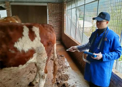
In recent years, the rise of portable ultrasound systems has revolutionized small animal practice. Handheld and wireless devices like the BXL-V50 have allowed general practitioners and mobile vets to bring high-resolution imaging directly to the patient, whether in an exam room, surgery suite, or even at home.
These devices offer key features:
-
Waterproof and rugged construction for daily clinical use
-
High-definition grayscale imaging for detailed soft tissue views
-
Long battery life—up to 7 hours—for mobile service use
-
USB and cloud storage for easy report sharing and archiving
Portability also improves workflow, especially in clinics that lack space for full ultrasound suites. More importantly, it allows timely intervention during emergencies like internal bleeding, urinary obstruction, or cardiac arrest.
Education and Training: The Key to Effective Use
In North America, organizations like the American College of Veterinary Radiology (ACVR) and the International Veterinary Ultrasound Society (IVUSS) offer courses and certifications. In the U.K., ultrasound training is often built into postgraduate Continuing Professional Development (CPD) programs.
Despite the technology being user-friendly, effective scanning requires skill. Learning probe positioning, image interpretation, and how to differentiate between normal and pathological findings takes time and practice.
Thankfully, many young veterinarians are graduating with at least basic ultrasound knowledge, and clinics are investing in staff training. Online modules and telemedicine-based scan reviews are also helping bridge the knowledge gap.
Common Limitations and How to Address Them
While ultrasound is invaluable, it does have limitations:
-
Operator-dependent: Image quality and interpretation vary with the user’s skill.
-
Limited depth penetration: Not ideal for visualizing lungs (unless fluid is present) or deep thoracic structures in large dogs.
-
Air and bone interference: Air in the bowel or overlying gas can obscure views, and bones cast shadows that limit visualization.
To work around these, experienced practitioners often:
-
Use fasting protocols to reduce gas in abdominal scans
-
Choose the correct probe frequency for different body sizes
Future Trends in Veterinary Ultrasound
Global demand for better, faster, and more affordable diagnostics has pushed ultrasound technology forward. Some notable trends:
-
AI integration: Artificial intelligence is being used to auto-detect structures and measure dimensions.
-
Teleultrasound: Vets in rural clinics can now upload scans for remote radiologist interpretation.
-
3D and elastography: New imaging modes are enhancing detail in liver and tumor assessments.
-
Wearable ultrasound: Under development for monitoring recovery or chronic disease in real time.
As technology becomes more affordable and intuitive, more general practitioners—not just specialists—will rely on ultrasound as a frontline tool.
Conclusion
From identifying abdominal masses to confirming pregnancies and evaluating heart function, ultrasound has transformed how we diagnose and treat small animals. Its non-invasive nature, real-time results, and expanding accessibility make it a must-have in modern veterinary practice.
As an experienced vet working in both first-opinion and referral settings, I’ve seen the difference ultrasound makes—not only in clinical outcomes but in client confidence and patient comfort. With the rise of compact, powerful systems like the BXL-V50 and continued advances in training and software, the future of small animal ultrasound looks brighter than ever.
For practitioners looking to upgrade their diagnostic tools or pet owners seeking the best care, embracing ultrasound means embracing precision, compassion, and progress in animal health.
References
-
Cornell University College of Veterinary Medicine. (2024). Diagnostic Imaging: The Role of Ultrasound in Small Animal Practice. https://www.vet.cornell.edu/hospitals/imaging
-
International Veterinary Ultrasound Society (IVUSS). (2023). Ultrasound Education and Certification for Vets. https://www.ivuss.org
-
British Small Animal Veterinary Association (BSAVA). (2024). Ultrasound Guidelines for Small Animal Practice. https://www.bsava.com/Resources/Veterinary-Resources/Ultrasound
-
American College of Veterinary Radiology (ACVR). (2023). Veterinary Diagnostic Imaging Standards. https://www.acvr.org
-
Veterinary Practice News. (2023). Ultrasound Technology Improves Diagnostics in Everyday Practice. https://www.veterinarypracticenews.com