How to Evaluate the Cattle Urinary Bladder Effectively on Ultrasound
Understanding the structure and health of the urinary bladder in cattle plays a critical role in diagnosing urogenital diseases, monitoring reproductive status, and guiding treatment decisions. Among the available diagnostic tools, ultrasonography stands out for its precision, non-invasiveness, and capacity to provide real-time imaging. For veterinarians and livestock producers in many parts of the world, ultrasound examination of the bladder has become an essential part of large animal diagnostics—especially in situations where symptoms like dysuria, hematuria, or distended abdomen appear.

Importance of Bladder Evaluation in Cattle
Cattle may present with a range of urinary system problems, including urolithiasis (urinary stones), bladder rupture, cystitis (bladder inflammation), and congenital malformations. While clinical signs such as straining to urinate, reduced urine flow, and abdominal swelling can offer clues, these are often non-specific. Ultrasound imaging fills the gap by offering direct visualization of bladder wall thickness, internal contents, and adjacent structures such as the ureters and reproductive organs.
In many Western veterinary settings, early and accurate bladder evaluation is a routine component of internal medicine and reproductive exams. From feedlot bulls with suspected obstruction to dairy cows suffering postpartum complications, ultrasonography helps clarify the clinical picture and enables veterinarians to make timely decisions.
Selecting the Right Equipment and Probe
Evaluating the bladder effectively depends heavily on using the right ultrasound equipment. A B-mode ultrasound machine with a convex or microconvex probe (3.5–5.0 MHz) is typically recommended for adult cattle. For calves or small-bodied breeds, higher-frequency linear probes (6.0–8.0 MHz) may provide greater resolution, especially for superficial scanning.
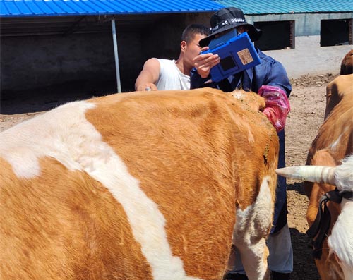
The probe is generally placed transabdominally in the caudal ventral abdomen, just cranial to the udder or scrotum, depending on the sex of the animal. In females, rectal ultrasonography using a linear probe may also offer a clear view of the bladder, particularly when evaluating in conjunction with the uterus or ovaries.

Technique: How to Perform a Proper Scan
A full bladder provides the best imaging window. If possible, scanning should be performed prior to urination to ensure optimal bladder distension. Here’s the basic scanning process used widely in veterinary practice:
-
Restrain the animal calmly, ideally in a chute or stocks.
-
Apply coupling gel liberally to the probe to maximize contact.
-
Place the probe transversely or longitudinally on the ventral abdomen near the pelvic brim.
-
Adjust depth and gain to focus on the bladder region.
-
Observe the bladder for:
-
Wall thickness and symmetry
-
Internal echogenic material (e.g., debris, blood clots, stones)
-
Free fluid around the bladder (which may indicate rupture)
-
In male cattle, the bladder often lies deeper and may require more scanning effort, particularly in bulls with large preputial or scrotal masses.
Normal Bladder Appearance on Ultrasound
In a healthy cow or bull, the bladder appears as a round to oval anechoic (black) structure with smooth, thin, echogenic walls. Its size varies significantly depending on hydration status and time since last urination.
-
Wall thickness should generally be less than 3 mm in a well-distended bladder.
-
Bladder contents should be uniformly anechoic unless there’s sediment, which may appear as echogenic (white) specks or floating debris.
One important ultrasonographic feature to consider is gravity-dependent sedimentation. While minor sediment is not uncommon, especially in animals on high-protein diets, large accumulations or mobile echogenic material could suggest cystitis or crystalluria.
Common Abnormal Findings and Their Interpretation
-
Thickened bladder wall (>5 mm): This may indicate chronic cystitis, which can arise from infection, trauma, or long-term catheterization.
-
Echogenic debris or clots: Blood clots, pus, or crystals within the bladder signal more severe inflammation or trauma, possibly secondary to urolithiasis.
-
Bladder stones (uroliths): These appear as hyperechoic (bright white) structures with strong distal acoustic shadowing. The location and mobility of stones are crucial for management—obstructive stones near the bladder neck often require surgical intervention.
-
Bladder rupture: In severe obstruction cases, the bladder may rupture, especially in young bulls. Free fluid (uroperitoneum) appears as anechoic spaces between loops of intestines or around organs. The bladder itself may be collapsed or difficult to visualize.
-
Congenital anomalies: Rare but sometimes encountered, conditions like bladder diverticula or ureteral ectopia may show up on ultrasound, often in conjunction with recurring urinary infections.
Ultrasonography in Reproductive vs. Urinary Diagnostics
In many cases, bladder imaging complements reproductive tract examinations, especially in females. The uterus, cervix, and bladder lie in close proximity, and during post-calving evaluations, retained placental fragments or uterine infections may mimic or mask bladder abnormalities. An experienced veterinarian can scan these regions sequentially to differentiate between the reproductive and urinary causes of illness.
For example, in postpartum cows with abdominal distension and foul-smelling discharge, ultrasound helps distinguish pyometra (uterine infection with pus accumulation) from bladder distension or rupture.
In bulls, evaluating the bladder is often paired with examination of the seminal vesicles and accessory sex glands, particularly in breeding soundness exams. The presence of bladder wall abnormalities or residual urine could suggest subclinical inflammation impacting reproductive performance.
The Global Veterinary Perspective
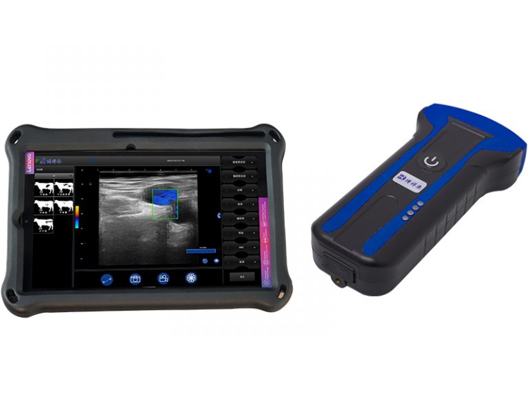
Across North America, Europe, and Australia, routine bladder scanning is now a standard part of bovine diagnostics, especially on dairy farms with high-producing cows prone to postpartum complications. In these regions, ultrasound machines like the BXL-V50—rugged, portable, and waterproof—are favored for fieldwork. These devices provide high-resolution images even in suboptimal conditions, with long battery life suited for remote or mobile practices.
In developing regions, the challenge often lies in access and training. However, NGOs and veterinary outreach programs have increasingly introduced portable ultrasound systems to rural veterinarians, improving the diagnosis of common urinary and reproductive disorders in both cattle and buffaloes.
Online veterinary courses and telemedicine platforms also help standardize knowledge. Resources like the University of Guelph’s “Dairy Health Management” program or the FAO’s guidelines on cattle health offer case-based examples of bladder pathology detected via ultrasound.

Clinical Applications and Decision-Making
Real-time bladder evaluation leads to faster and more accurate decisions in several scenarios:
-
Urethral obstruction: Rapid identification of bladder distension confirms the need for immediate intervention.
-
Bladder rupture: Avoids unnecessary medical treatment and guides emergency surgery or euthanasia.
-
Cystitis or infection: Detecting bladder wall thickening or debris supports the use of antimicrobials.
-
Monitoring recovery: Follow-up scans track healing after catheterization or surgery.
Moreover, pre-purchase evaluations in breeding bulls sometimes include bladder scans, especially in regions where urolithiasis is endemic. Early detection of sediment or early-stage inflammation can prevent the sale of animals with poor reproductive outlook.
Challenges and Limitations
While ultrasonography is a powerful tool, it has limitations:
-
Bladder scanning in large, overweight bulls can be technically difficult due to fat layers or deep positioning.
-
Collapsed or empty bladders provide poor imaging windows, requiring strategic timing of the scan.
-
Artifacts like gas shadowing from intestines may obscure views, necessitating repositioning or re-imaging.
Also, ultrasound cannot replace biochemical analysis. In some cases, urinalysis or abdominocentesis is still required to confirm findings, such as when differentiating between infectious cystitis and sterile inflammation.
Final Thoughts
Ultrasonography is now indispensable in modern cattle practice, particularly when evaluating the urinary bladder. With the right technique and equipment, veterinarians can identify subtle changes in bladder anatomy and content that would otherwise go unnoticed. In doing so, they not only improve diagnostic accuracy but also reduce suffering and enhance outcomes for the animals.
In countries where ultrasound adoption is widespread, bladder evaluation has moved from a niche skill to a routine check—just as essential as checking rumen fill or reproductive status. As technology becomes more affordable and user-friendly, more farmers and field veterinarians worldwide are embracing its use, resulting in better cattle care, quicker interventions, and fewer costly surprises.
Reference Sources:
-
Whitaker, D. A., & Smith, E. (2021). Veterinary Ultrasonography in Food-Producing Animals. Journal of Veterinary Imaging.
-
FAO. (2023). Field Diagnosis of Cattle Diseases. https://www.fao.org/3/ah847e/ah847e00.htm
-
Beef Cattle Institute. (2023). Use of Ultrasound for Diagnostic Evaluation in Cattle. https://www.beefcattleinstitute.org/ultrasound-diagnosis
-
University of Guelph – Dairy Health Management Program. https://dairyhealth.uoguelph.ca


