How Farms Detect Horse Tendon Tears with Ultrasound
Tendon injuries in horses can be heartbreaking—for both the animal and the people who care for them. Whether it’s a racehorse coming off a winning streak or a quiet trail horse who suddenly starts limping, tendon tears are common, painful, and can take months (or even years) to heal properly. For years, detecting and managing these injuries was mostly based on physical examination, swelling, heat, and guesswork. But now, thanks to modern veterinary ultrasound, farms can get an accurate, real-time view of what’s happening inside a horse’s leg without any invasive procedures.

Why Tendon Tears Are So Tricky
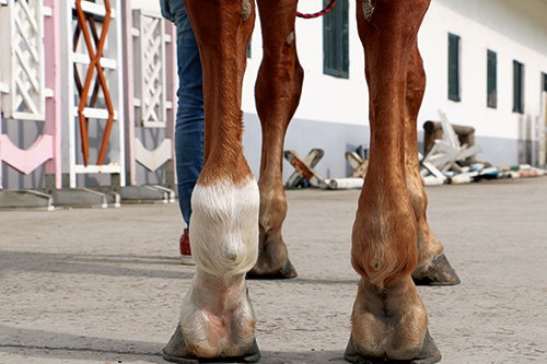
Tendons in horses—especially the superficial digital flexor tendon (SDFT) and deep digital flexor tendon (DDFT)—play a crucial role in movement and weight-bearing. When these tendons get overstretched or torn due to fatigue, poor footing, or repetitive strain, horses can become lame almost immediately. The tricky part? Not all tears are visible on the surface. Some horses may just show mild swelling or intermittent lameness, making diagnosis difficult without imaging.
Many horse owners used to rely on the “poke and feel” method—pressing along the back of the leg to check for swelling or heat. But this method isn’t reliable enough, especially in early stages or partial tears. That’s where ultrasound truly shines.
What Ultrasound Actually Shows in a Horse’s Leg
When a horse injures a tendon, the structure of the tendon fiber becomes disrupted. Instead of neatly aligned parallel fibers, there may be gaps, fluid, or scar tissue forming. A portable veterinary ultrasound can pick up all these signs within minutes.
Using a linear probe with high frequency (usually between 7.5 to 12 MHz), the vet applies gel on the horse’s leg and slowly scans along the tendon. Healthy tendons appear as homogenous, tightly packed fibrillar patterns on the ultrasound image. But when there’s a tear, you’ll often see hypoechoic (darker) regions, indicating fluid or disrupted tissue.
Experienced equine vets can even grade the severity of the injury on the spot—mild lesions with small fiber disruption, moderate tears with partial separation, and severe injuries with complete rupture.
Real-Life Use on Farms: How It’s Done
Let’s say you’re on a breeding or training farm with performance horses. A gelding starts showing slight lameness after a workout. There’s no obvious swelling, and the farrier didn’t notice anything off with the hoof. Before ultrasound, you’d probably rest the horse for a couple of weeks, hoping for improvement. But now, many farms have access to a mobile vet with an ultrasound machine—or even their own portable units.
A quick scan on the back of the cannon bone can rule out or confirm tendon injury. If there’s a tear, the scan gives the exact size, location, and nature of the damage, which is vital for building a rehab plan. Many farms use serial ultrasounds—scanning every few weeks—to track healing and adjust training accordingly. This prevents pushing a horse too hard, too soon, which is one of the top causes of reinjury.
Not Just for Diagnosis: Monitoring Healing Too
One of the most misunderstood things about tendon injuries is that they often “look” healed before they actually are. The swelling goes down, the horse walks soundly, but internally, the tissue may still be weak and fragile.
This is where ultrasound makes all the difference. By visually assessing how the tendon fibers are re-aligning and whether scar tissue is forming properly, vets can make informed decisions on when to increase exercise or return to light work. On many U.S. and European farms, a horse isn’t cleared to go back under saddle until the ultrasound shows sufficient healing—not just visual or behavioral improvement.
Early Detection = Better Prognosis
In top-level racing and sport horse operations, ultrasound isn’t just a response tool—it’s also a preventive one. Regular scans, especially during intense training seasons, help catch microtears or early fiber disruption before they become full-blown ruptures.
These early lesions might not cause obvious lameness but can be picked up as slight changes in echogenicity or fiber alignment. Catching them early allows trainers to reduce workload, apply supportive therapies (like stem cells, PRP, or shockwave), and avoid career-ending injuries.
Common Tendon Injuries Diagnosed by Ultrasound
-
SDFT core lesions – The most common in racehorses. Easily seen as a central dark area.
-
DDFT tears – Often occur within the digital sheath or inside the hoof, requiring a special probe or transducer angle.
-
Suspensory ligament desmitis – Especially in sport horses, this ligament injury is tricky to detect without imaging.
-
Tendon sheath effusion or infection – Shows up as excess fluid or abnormal synovial structures on ultrasound.
International Practices and Equipment Trends
In the U.S., U.K., Australia, and New Zealand—regions with robust equine industries—portable ultrasound machines are now standard on most veterinary trucks. Many large equine clinics use Doppler or even 3D ultrasound to assess blood flow and tissue texture. While B-mode ultrasound (2D grayscale) remains the workhorse for tendon evaluation, color Doppler helps assess inflammation and vascularity during healing.
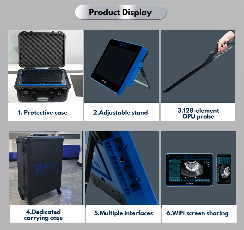
Some farms have trained their barn managers or senior grooms to operate basic ultrasound scans, especially for follow-up monitoring. Devices like the Butterfly iQ+ Vet or the BXL-S300 have made it easier than ever for farms to bring imaging in-house.
How Ultrasound Changed Recovery Plans
Before ultrasound became widespread, most tendon rehab plans were based on time—six months of rest, then gradual reintroduction to work. But tendons don’t always heal on schedule. Now, with ultrasound-guided rehab, rest can be adjusted precisely to the actual condition.
This saves time in some cases (when healing is faster) and prevents disaster in others (when scarring is incomplete or improperly aligned). Horses with ultrasound-guided treatment also tend to have lower reinjury rates and better long-term outcomes.
Beyond Imaging: A Communication Tool
Farmers and trainers also find that ultrasound images help with communication. A printed scan showing a clear lesion allows vets to explain exactly what’s happening, and owners are more likely to follow rest and rehab plans when they see the damage. It’s a practical education tool that improves compliance and decision-making.
Conclusion
When it comes to managing tendon injuries in horses, ultrasound has become one of the most powerful tools on the farm. It takes the guesswork out of diagnosis, helps track healing, prevents reinjury, and ultimately keeps horses sounder, longer.
From top-level racehorses to family ponies, the ability to “see inside” a leg in real-time changes everything. As more farms around the world adopt this technology, the days of waiting, guessing, and hoping for healing are becoming a thing of the past.


