Assessing Backfat Thickness in Swine Using Handheld Ultrasound Devices
Accurately managing fat deposition in pigs is one of the key strategies for optimizing meat quality, feed efficiency, and overall profitability in swine production. Among the many modern technologies aiding this effort, handheld ultrasound devices stand out as a practical, non-invasive, and real-time method for assessing backfat thickness. This technique is increasingly being adopted by pig farmers around the world—not only in large-scale operations but also among smallholder farms seeking greater precision in breeding and nutrition management.
In this article, I’ll explain how we use portable ultrasound tools to evaluate backfat in pigs, explore the biological principles behind fat deposition, and share practical insights from both my experience and global perspectives in swine management.
Why Measure Backfat in Swine?
Backfat thickness is a reliable indicator of body condition and energy reserves in pigs. Monitoring this trait is essential in various production phases:
-
Breeding sows: Maintaining optimal backfat helps ensure good reproductive performance, prevents over-conditioning, and reduces risks during farrowing.
-
Growing-finishing pigs: Ensures ideal carcass composition, improves feed conversion ratio (FCR), and maximizes meat yield.
-
Boars: Helps maintain optimal weight and health for sperm quality and longevity.
Handheld ultrasound enables precise and repeatable measurement of subcutaneous fat at key anatomical sites, such as the last rib or P2 position (6.5 cm from the midline), providing crucial data for informed decisions.
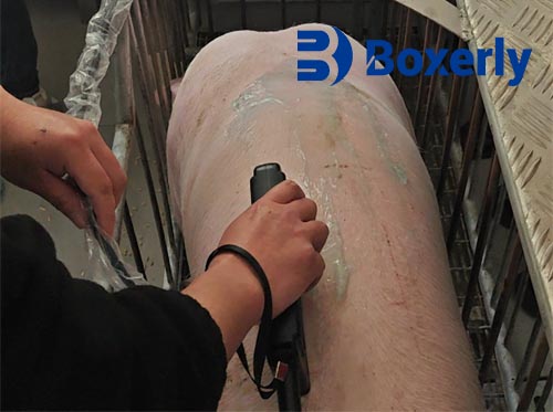
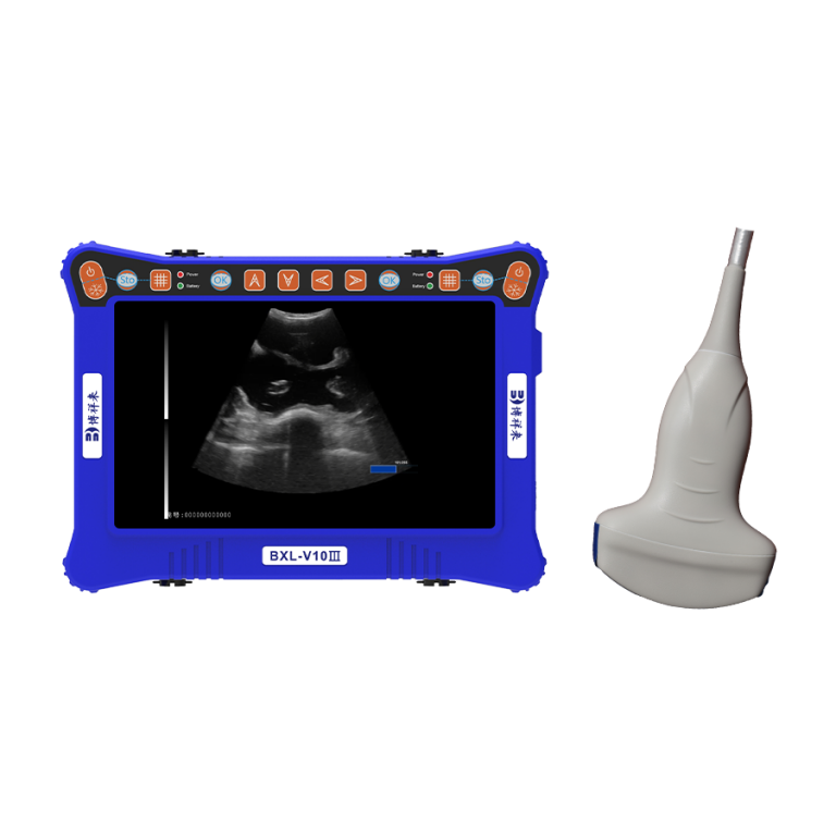
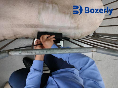
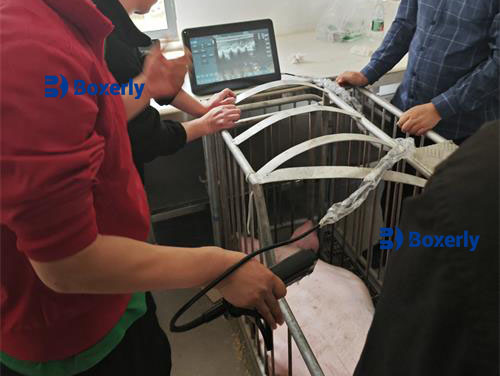
How Handheld Ultrasound Works in Practice
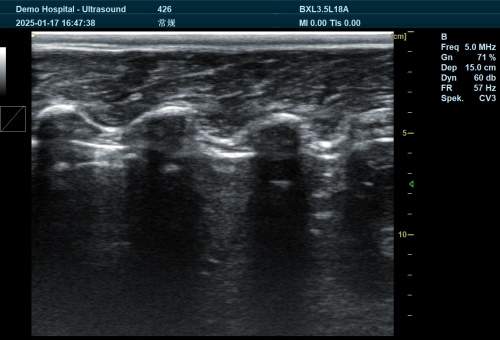
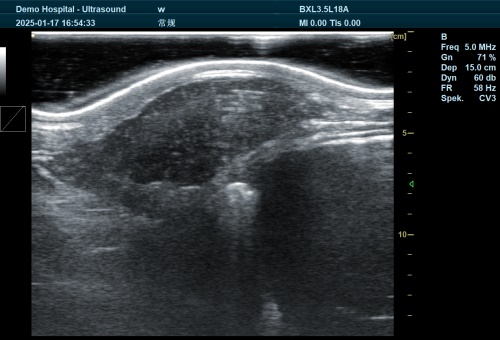
Handheld ultrasound devices typically use B-mode (brightness mode) imaging to capture cross-sectional visuals of tissue layers. In pigs, the most common application is measuring the distance between the skin surface and the top of the Longissimus dorsi muscle—this gap represents backfat thickness.
The process involves:
-
Restraint and positioning: The pig is secured safely, and the hair may be clipped for better contact.
-
Applying ultrasound gel: Improves signal transmission between the probe and skin.
-
Scanning: The probe is placed at standard landmarks such as the P2 site.
-
Image analysis: The user identifies skin, fat, and muscle boundaries to measure backfat.
Advanced devices offer image freezing, measurement tools, and even automatic analysis software, streamlining the evaluation.
Biological Patterns of Fat Deposition in Pigs
Pigs deposit fat in a specific biological order that reflects their developmental stage:
-
Internal (visceral) fat
-
Subcutaneous fat (backfat)
-
Intermuscular fat
-
Intramuscular fat (marbling)
Subcutaneous fat, the target of most routine backfat measurements, develops as pigs approach market weight. Handheld ultrasound helps detect this growth phase early and track its rate over time.
Foreign research confirms this sequence. For example, a study by McLaren et al. (2019) in the Journal of Animal Science demonstrated that backfat development in crossbred pigs follows a predictable curve, with rapid growth between 60–100 kg live weight and a plateau afterward. Recognizing this curve enables farmers to time feed adjustments, breeding, or slaughter more accurately.
Global Use and Research on Swine Ultrasound
In countries like the United States, Canada, and Denmark, ultrasound use in pig farming is integrated into national genetics and carcass grading systems. Portable ultrasound is often used by swine consultants and AI technicians to record backfat data, which is then fed into breeding databases to estimate heritability and genetic merit.
A 2022 Danish study published in Livestock Science found that routine backfat assessment using ultrasound reduced sow mortality and improved litter performance over three breeding cycles. The key takeaway was that handheld imaging, when used consistently, leads to better-managed body condition and fewer reproductive issues.
Advantages of Using Handheld Ultrasound on the Farm
✔ Non-Invasive and Animal-Friendly
There’s no need for sedation or blood sampling. Ultrasound causes minimal stress to the pig, allowing frequent measurements.
✔ Real-Time Feedback
Farmers get immediate visual and numerical data, enabling on-the-spot decisions regarding nutrition and management.
✔ Portability
Devices are compact and easy to carry—suitable for both indoor and outdoor units.
✔ Economic Benefits
Accurate body condition assessment leads to more efficient feed use, reduced waste, and improved reproductive performance.
✔ Breeding Optimization
By selecting pigs with ideal backfat, farmers can breed for both leanness and fertility—two critical economic traits.
Common Parameters Measured by Ultrasound in Swine
| Parameter | Purpose |
|---|---|
| Backfat Thickness (P2) | Indicator of body condition and fatness |
| Loin Eye Area (LEA) | Reflects muscle development and meat yield |
| Intramuscular Fat Estimate | Suggests marbling and meat quality |
| Muscle Depth | Helps differentiate lean from fatty carcasses |
Some high-end handheld scanners offer dual-mode functionality—allowing both backfat and muscle area measurements with one device.
Integrating Ultrasound into Management Decisions
Handheld ultrasound devices are most useful when integrated into a regular farm protocol. For example:
-
Before breeding: Avoid inseminating under-conditioned gilts or over-fat sows, both of which risk poor performance.
-
During gestation: Adjust feed based on real-time body condition to avoid excessive weight gain or loss.
-
Before slaughter: Ensure optimal market weight and fatness for premium carcass grading.
On my farm, ultrasound checks every two weeks during key production periods have allowed us to reduce culling rates, optimize feed rations, and improve carcass consistency.
Breed and Diet Influence on Backfat Development
Different breeds show varying fat deposition tendencies. For instance:
-
Duroc pigs: Tend to deposit more backfat but also deliver high marbling.
-
Landrace and Yorkshire: Leaner, with slower backfat accumulation.
Diet also plays a vital role:
-
High-energy diets: Promote faster fat deposition, especially in late-finishing pigs.
-
Controlled energy rations: Useful in gestation to prevent over-conditioning.
By adjusting nutrition based on ultrasound findings, we align feed efficiency with growth targets and meat quality goals.
Limitations and Considerations
While ultrasound is a powerful tool, it’s not without its challenges:
-
Training required: Accurate image interpretation demands practice.
-
Environmental interference: Excessive dirt, hair, or wet skin may affect image clarity.
-
Cost barrier: Initial investment may be high, though long-term benefits usually outweigh this.
Still, many countries offer extension training or cooperative equipment programs to help smaller farms adopt this technology.
Future of Handheld Ultrasound in Swine Production
As device prices drop and software improves, more farmers are incorporating ultrasound into regular herd monitoring. Emerging innovations include:
-
AI-assisted image analysis: Automatically detects and labels fat/muscle layers.
-
Wireless probes and smartphone apps: Offer data logging and sharing features.
-
Integration with farm software: Allows long-term tracking of individual animals.
The future lies in data-driven pig farming, where every management decision is backed by real-time imaging and analytics.
Conclusion
Using handheld ultrasound to assess backfat thickness in pigs is no longer just a high-tech luxury—it’s becoming a practical necessity. From breeding and feeding to finishing and slaughter, this tool empowers swine producers to make more informed, accurate, and profitable decisions.
On my farm and in many operations worldwide, the routine use of ultrasound has led to better body condition control, reduced feed waste, enhanced reproductive outcomes, and more uniform meat quality. Whether you’re managing 50 pigs or 5,000, this technology provides a clear, objective picture of what’s happening beneath the skin—so you can manage smarter, not harder.
References
-
McLaren, D. G., et al. (2019). Ultrasound Measurement of Fat and Muscle in Swine. Journal of Animal Science. https://doi.org/10.1093/jas/skz082
-
Hansen, M. L., & Pedersen, H. J. (2022). Body Condition Management in Danish Sows Using Ultrasound. Livestock Science. https://www.sciencedirect.com/science/article/pii/S1871141322000123
-
National Swine Improvement Federation. (2021). Guidelines for Ultrasound in Swine Genetic Evaluation. https://www.nsif.com