Understanding How Veterinary Ultrasound Works in Animal Diagnosis
Veterinary ultrasound has become an indispensable tool in modern animal health management. Whether used on a sheep farm to diagnose pregnancy, in a dairy barn to monitor ovarian activity in cows, or in a veterinary clinic to assess abdominal structures in dogs and cats, ultrasound technology offers a non-invasive, real-time window into the animal body. However, understanding how ultrasound works is crucial to using it effectively.
In this article, we’ll explore the basic principles of animal ultrasound imaging, also known as ultrasonography. We’ll cover how ultrasound travels through body tissues, how echoes are formed, how images are generated, and what factors influence the clarity and accuracy of the results. Along the way, we’ll compare these insights with widely accepted practices in veterinary medicine globally.
How Ultrasound Waves Travel in the Animal Body
At its core, ultrasound is a form of mechanical sound wave that travels through a medium—in this case, animal tissues. Unlike X-rays that use ionizing radiation, ultrasound is based on high-frequency sound waves (usually 2 to 15 MHz) that are above the range of human hearing.
Ultrasound waves travel in straight lines and have good directionality. This property allows veterinary technicians or veterinarians to target specific organs or tissue layers during examination. When these waves encounter a boundary between two tissues with different acoustic impedances (for example, between muscle and fat, or between fluid and tissue), part of the wave is reflected back toward the probe—this reflected wave is called an echo—while the rest may be refracted or absorbed as it continues through the next tissue layer.
Reflection, Refraction, and Attenuation: The Physics Behind the Images
When ultrasound waves strike a large boundary between tissues of significantly different densities (like between fluid and bone), strong echoes are produced. These echoes travel back to the transducer (probe), where they are converted into electrical signals and then displayed as bright spots on the monitor. The greater the difference in acoustic impedance, the stronger the echo and the brighter the spot.
However, not all boundaries are large. If the interface is smaller than the wavelength of the ultrasound wave (as in capillaries or small tissue nodules), scattering occurs. This causes the energy to disperse in multiple directions, often leading to weaker and less focused echoes.
Additionally, as ultrasound travels through tissues, it undergoes attenuation—a gradual decrease in intensity due to absorption, scattering, and reflection. The further the wave travels, the weaker it becomes. This is why deeper structures often appear darker or less distinct on the sonogram.
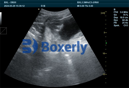
The Concept of B-Mode (Brightness Mode) Imaging
In veterinary medicine, B-mode ultrasonography (Brightness mode) is the most commonly used format. This real-time imaging technique displays a two-dimensional cross-sectional image of the organ being scanned. Each echo is represented as a dot on the screen, with its brightness corresponding to the strength of the echo.
No echo results in a black area (anechoic), weak echoes appear gray (hypoechoic), and strong echoes appear white (hyperechoic). For example, fluid-filled structures like the bladder or amniotic sac are usually black because they don’t reflect sound waves well. In contrast, bones or fibrous tissues appear bright white.
This grayscale imaging allows veterinarians to assess the size, shape, texture, and relative position of organs and abnormalities. It is particularly useful in reproductive management, abdominal diagnostics, cardiac assessments, and even in identifying tumors or foreign bodies.
From Point to Picture: Creating the Sonogram
The image produced on the ultrasound monitor is called a sonogram. It is essentially a map of returning echoes arranged according to their origin and intensity. These echoes are generated in rapid succession—thousands of times per second—as the probe emits and receives sound waves.
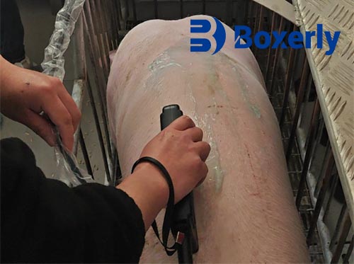
To build a comprehensive image, the probe must be moved or rotated to scan the organ from multiple angles. Common probe manipulation techniques include:
-
Linear movement (sliding the probe in a straight line)
-
Fan movement (pivoting the probe in a fixed spot like a windshield wiper)
-
Rocking motion (tilting the probe toward and away from the operator)
-
Pressure variation (pressing gently to displace superficial tissue or fluid)
These techniques help create a detailed and accurate depiction of the organ under study. For larger animals or deeper tissues, lower-frequency probes (e.g., 3.5 MHz) are used, while for smaller animals or superficial structures, higher-frequency probes (e.g., 7.5 to 10 MHz) provide better resolution.
Ultrasound Image Interpretation: Density and Contrast
Different tissues in the animal body absorb and reflect ultrasound waves differently based on their density and composition. Here’s how typical structures appear on an ultrasound image:
-
Fluid (e.g., bladder, cysts, amniotic fluid): Anechoic (black)
-
Fat: Hypoechoic (dark gray)
-
Muscle: Medium echogenicity (gray)
-
Fibrous tissue or connective tissue: Hyperechoic (white)
-
Gas or air (e.g., lungs, intestines): Highly reflective but scatter-prone, producing artifacts
-
Bone: Very hyperechoic, often casting a shadow beyond it (due to total reflection)
Understanding these patterns allows veterinarians to distinguish between normal and abnormal findings and to track changes in the body over time.
Why Real-Time Imaging Matters
The real-time aspect of B-mode ultrasound is one of its greatest advantages. Unlike static imaging methods, ultrasound provides continuous feedback as the operator moves the probe. This is essential when scanning dynamic organs like the heart, uterus during pregnancy, or gastrointestinal system.
For example, in reproductive management of dairy cows or sheep, veterinarians can use real-time ultrasound to detect ovulation, monitor embryo development, or confirm fetal heartbeat. These real-time updates also allow immediate adjustments during the scan, such as changing the probe angle or adjusting depth and gain settings on the machine.
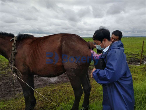
Global Perspective on Veterinary Ultrasonography
Veterinary professionals around the world—whether in North America, Europe, or Asia—use ultrasound as a front-line diagnostic tool due to its non-invasive nature, portability, and versatility. In many developing regions, mobile ultrasound units are transforming livestock health management by enabling early diagnosis and timely intervention.
Moreover, with increasing adoption of digital technologies, modern ultrasound machines can now store images, export video clips, and even integrate with herd management software for record-keeping and analysis. In research settings, ultrasound is being used to evaluate muscle growth, fat deposition, and internal pathology in ways that were not possible two decades ago.
Challenges and Considerations
Despite its many benefits, ultrasound imaging is not without limitations. The quality of the image depends on multiple factors:
-
Operator skill and experience
-
Quality and calibration of the machine
-
Frequency and type of probe
-
Animal size, species, and body condition
-
Presence of gas, feces, or excessive fat that may block sound waves
Thus, continuous training and equipment maintenance are essential for ensuring accurate diagnosis.
Conclusion
Understanding how veterinary ultrasound works is the first step toward making the most of this powerful tool. From wave transmission and echo formation to image interpretation and real-time diagnosis, each aspect of ultrasonography is grounded in physics yet designed for practical use.
As global livestock industries grow more reliant on precision diagnostics, ultrasound stands out as a cost-effective, safe, and informative solution. Whether you’re a farm manager, a veterinary student, or an experienced practitioner, mastering the basic principles of animal ultrasound will undoubtedly enhance your ability to deliver better animal care.