Luteal Blood Flow Ultrasound: Understanding Corpus Luteum Vascularization in Cattle Reproduction

Luteal Blood Flow Ultrasound: Understanding Corpus Luteum Vascularization in Cattle Reproduction
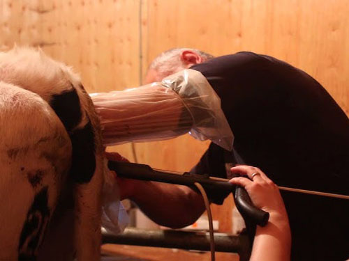
Accurate reproductive monitoring is essential for improving fertility and breeding efficiency in modern dairy and beef cattle operations. Among the different reproductive examination techniques, luteal blood flow ultrasound has become an increasingly valuable application of veterinary Doppler imaging.
The corpus luteum (CL) is a temporary ovarian structure that develops after ovulation and plays a critical role in progesterone production. Its development and functional activity are closely associated with changes in blood circulation.
Using Color Doppler and Power Doppler ultrasound, veterinarians can visualize blood-flow signals within the corpus luteum and obtain additional information about ovarian vascularization.
Unlike conventional grayscale ultrasound, Doppler imaging provides functional information about blood movement, helping veterinarians and reproductive specialists evaluate luteal development and ovarian conditions.
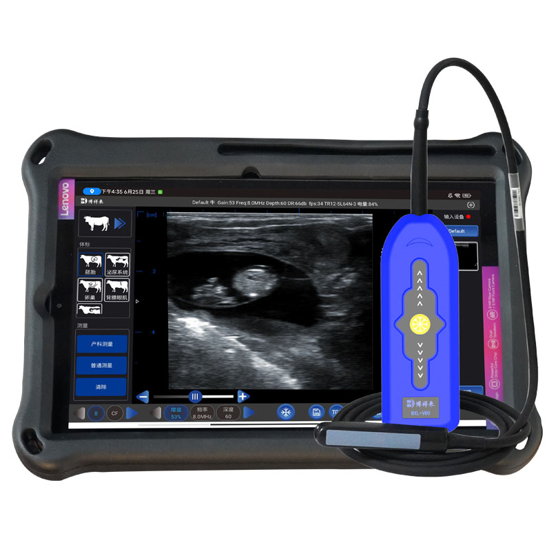
BOXERLY, a professional veterinary ultrasound manufacturer from China, provides wireless color Doppler ultrasound systems designed for advanced cattle reproductive examinations.
1. What Is Luteal Blood Flow Ultrasound?
Luteal blood flow ultrasound is an examination technique used to evaluate blood circulation within the corpus luteum.
After ovulation, the remaining follicular cells undergo structural changes and develop into the corpus luteum. During this process, a new network of blood vessels forms to support the metabolic and endocrine activities of the developing structure.
Blood circulation supplies oxygen and nutrients and supports the delivery of substances involved in progesterone production.
Veterinarians can use Doppler ultrasound to visualize blood-flow signals in the corpus luteum and examine its vascular distribution.
During a luteal blood flow examination, several characteristics may be evaluated:
Distribution of detectable blood-flow signals.
Relative vascularization within the corpus luteum.
Changes in luteal blood-flow patterns over time.
Differences in vascular signals during luteal development and regression.
Relationship between luteal vascularization and reproductive status.
This examination provides additional functional information that cannot be obtained from conventional B-mode ultrasound alone.
2. Why Is Luteal Blood Flow Important in Cattle?
The corpus luteum is essential for maintaining the normal reproductive cycle and supporting early pregnancy in cows.
Its primary endocrine function is progesterone secretion.
Progesterone helps prepare the uterus for pregnancy and supports the physiological environment required for embryonic development.
Adequate blood circulation is important because the corpus luteum requires continuous delivery of oxygen, nutrients, and other substances necessary for hormone production.
Changes in luteal blood flow are therefore associated with changes in ovarian function.
Corpus Luteum Formation
Following ovulation, the corpus luteum develops rapidly. Its vascular network expands to support the increasing metabolic demands of luteal tissue.
Doppler ultrasound can help visualize the development of vascular signals during this process.
Luteal Functional Activity
A well-developed corpus luteum generally has substantial vascular support.
Evaluating its blood-flow signals provides additional information about luteal vascularization and can support the assessment of ovarian function.
Corpus Luteum Regression
When a cow is not pregnant, the corpus luteum normally undergoes regression at the end of the estrous cycle.
This process involves changes in luteal function, tissue structure, and blood circulation.
Serial Doppler examinations can help researchers observe these changes.
Early Pregnancy Support
In pregnant cows, maintaining corpus luteum function is essential for progesterone production.
Luteal blood-flow assessment can provide useful supplementary information during early pregnancy research and reproductive monitoring.
However, blood-flow signals alone cannot confirm pregnancy or determine pregnancy viability.
3. How Does Doppler Ultrasound Evaluate Luteal Blood Flow?
Doppler ultrasound uses changes in the frequency of returning ultrasound echoes to evaluate moving blood cells.
The ultrasound system processes these signals and displays blood-flow information in different visual formats.
For cattle reproductive examinations, Color Doppler and Power Doppler are particularly useful.
Color Doppler Ultrasound
Color Doppler displays blood-flow information as color signals superimposed on the grayscale image.
It provides information about flow direction and relative velocity, according to the selected color map and Doppler settings.
During corpus luteum examination, veterinarians can observe the distribution of blood-flow signals within and around luteal tissue.