Exploring Gastrointestinal Motility in Horse Using Portable Ultrasound Under Field Conditions
In modern equine veterinary medicine, the assessment of gastrointestinal (GI) motility is a vital aspect of diagnosing and managing digestive disorders, particularly colic—the leading cause of emergency interventions in horses. Traditionally, such evaluations were reliant on auscultation, palpation, and clinical signs. However, advancements in portable ultrasound technology have enabled more accurate, non-invasive, and real-time visualization of GI structures and their motility, even under field conditions. This article explores how veterinarians and equine practitioners in various parts of the world are leveraging portable ultrasound systems to monitor GI motility in horses, highlighting its advantages, limitations, and implications for field-based equine care.
Understanding Gastrointestinal Motility in Horses
Gastrointestinal motility refers to the coordinated contractions of the smooth muscles in the horse’s digestive tract, responsible for moving ingested material through the stomach, small intestine, cecum, and colon. Normal motility patterns are essential for digestion, nutrient absorption, and waste elimination. Disturbances in motility, whether due to inflammation, impaction, gas accumulation, or torsion, often lead to gastrointestinal discomfort and colic.
In field settings—especially during emergency farm calls or at equestrian events—quick and accurate assessment of GI motility can make a significant difference in treatment outcomes. The ability to perform this assessment without the need for referral to a hospital setting is especially valuable in rural or resource-limited environments.
Why Portable Ultrasound?
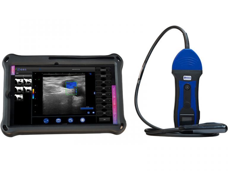
Portable ultrasound devices have revolutionized equine diagnostics in the field. Compact, battery-powered units with high-resolution imaging capabilities allow veterinarians to bring sophisticated diagnostics directly to the patient. Unlike radiography or endoscopy, ultrasound can be used without sedation, radiation exposure, or specialized infrastructure.
Key reasons for using portable ultrasound to assess equine GI motility include:
-
Non-invasiveness: There is no need for surgical exploration or sedation.
-
Real-time imaging: Motility can be observed as it happens, which is critical for dynamic evaluations.
-
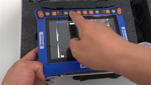
High portability: Light, durable units such as the BXL-V50 (although this article does not specifically promote any product) offer flexibility for fieldwork.
-
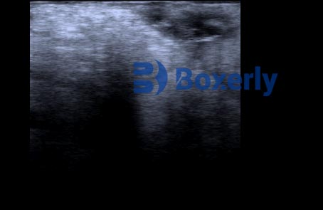
Diagnostic value: Helps distinguish between types of colic (e.g., gas, impaction, displacement), identify gut distension, monitor peristalsis, and detect fluid accumulation or wall thickening.
Common Ultrasound Sites for Assessing GI Motility
Ultrasound examination of the horse’s abdomen typically involves evaluating multiple windows along both flanks and ventral abdomen. Commonly assessed segments include:
-
Duodenum and jejunum (right side): Easily accessible and vital for observing small intestinal motility. Hyper- or hypomotility can be significant diagnostic indicators.
-
Cecum and right colon (right side): Normal motility appears as slow, large contractions.
-
Left colon and pelvic flexure (left side): These areas are prone to impaction and displacement.
-
Stomach and spleen (left side): Occasionally used for locating gas distension or foreign material.
A skilled veterinarian can scan these areas in under 10 minutes, making it a quick and effective tool for emergency evaluation.
What Foreign Practitioners Observe in the Field
In countries like the United States, United Kingdom, and Australia, equine veterinarians have been at the forefront of applying portable ultrasound in the field. A 2021 survey published in the Equine Veterinary Journal indicated that over 65% of equine veterinarians in rural practice use portable ultrasound routinely for gastrointestinal evaluations. These practitioners report several key observations:
-
Correlation with clinical signs: Decreased or absent motility often coincides with elevated heart rate, dry mucous membranes, and increased abdominal girth.
-
Motility scoring systems: Some vets use semi-quantitative scoring of contractions (e.g., <1 contraction per minute = hypomotile) to guide therapy.
-
Early decision-making: Visual confirmation of a non-motile bowel can justify early referral for surgery, even before rectal palpation or bloodwork.
-
Monitoring recovery: Post-surgical patients or those on medical management can be monitored non-invasively to track improvements.
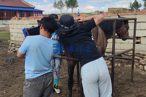
Techniques and Considerations in the Field
Conducting ultrasound in the field requires adaptations compared to hospital settings. Environmental light, wind, and animal movement all affect imaging quality. Based on field reports and clinical best practices, the following tips enhance success:
-
Clipping the hair is essential for optimal probe contact, even in cold weather.
-
Alcohol or ultrasound gel should be used generously to eliminate air interference.
-
Patient restraint (halter and lead) is often sufficient without sedation.
-
Battery-powered machines with long operational time (5–7 hours) and high screen brightness are preferred.
Modern machines offer multiple probes (convex, linear) suitable for different abdominal depths, with some devices featuring sun visors or touch screens for better visibility outdoors.
Clinical Applications and Case Insights
In international equine practice, GI ultrasound has become a frontline tool in diagnosing colic and evaluating treatment response. Below are two real-life case insights adapted from peer-reviewed reports and practitioner experiences:
Case 1: Small Intestinal Hypomotility in a Racehorse (USA)
A 5-year-old Thoroughbred presented with mild colic and reduced manure output. On-field ultrasound revealed significantly distended small intestine loops (>5 cm diameter) with no peristaltic movement observed over 3 minutes. Early referral and surgery confirmed a strangulating lipoma. The horse recovered after resection.
Case 2: Impaction Colic in a Draft Horse (UK)
A Belgian Draft gelding showed signs of abdominal discomfort. Field ultrasound displayed normal small intestinal motility but an enlarged left colon with minimal movement and hyperechoic content. The veterinarian opted for conservative medical treatment, including fluids and laxatives. Follow-up ultrasound showed progressive return of motility and reduced distension.
These cases underline the value of timely ultrasound-based assessment in guiding clinical decisions and improving survival rates.
Limitations and Challenges
Despite its many benefits, using ultrasound to evaluate GI motility has limitations:
-
Operator dependency: Experience plays a major role in image interpretation.
-
Intermittent motility: In normal horses, motility is not constant, so timing and patience matter.
-
Anatomical variation: Gas pockets can obscure deeper structures, especially in the large colon.
Nevertheless, when used by trained professionals, portable ultrasound serves as a reliable adjunct to clinical examination, rectal palpation, and laboratory testing.
International Research and Future Directions
Equine veterinary research continues to refine the role of ultrasound in gastrointestinal diagnostics. Some of the latest trends include:
-
Quantitative motility software: Algorithms that count contractions automatically are being tested.
-
Telemedicine integration: Images captured in the field can be transmitted to specialists for real-time consultation.
-
3D ultrasound and elastography: These may one day provide even deeper insights into tissue function and elasticity.
In the future, we may see portable ultrasound combined with AI-driven diagnostic models to further improve diagnostic accuracy in equine GI disorders.
Conclusion
The use of portable ultrasound to assess gastrointestinal motility in horses under field conditions has transformed equine veterinary practice. This non-invasive, real-time technique offers critical diagnostic insights that guide early intervention, improve prognosis, and reduce unnecessary surgeries. As field-ready ultrasound devices become more advanced and affordable, their adoption is expected to rise across the globe, particularly in rural and developing regions.
For equine practitioners and horse owners alike, understanding the power of ultrasound imaging in monitoring gut motility is crucial to ensuring timely, accurate care. Whether it’s identifying subtle signs of impaction or confirming the need for urgent referral, portable ultrasound is a game-changer in the field-based management of equine digestive health.
References
-
Freeman, D. E., & Schambourg, A. (2021). Equine Abdominal Ultrasonography in the Field. Equine Veterinary Education, 33(6), 281–289.
-
Corley, K. T. T., & Stephen, J. E. (2018). The Equine Hospital Manual. Wiley-Blackwell.
-
Rantanen, N. W. (2020). Diagnostic Ultrasonography in the Horse. Veterinary Clinics of North America: Equine Practice, 36(3), 435–452.
-
Equine Veterinary Journal (2023). Survey on Portable Ultrasound Use in Equine Practice.