How to Determine the Sex of a Cow’s Fetus by Ultrasound
Determining the sex of a cow’s fetus is an important aspect of modern cattle management, particularly in beef and dairy production. With the help of veterinary ultrasonography—especially real-time B-mode ultrasound—producers can obtain valuable information about the fetus as early as 55 to 60 days into pregnancy. This information helps optimize breeding strategies, improve herd structure, and enhance economic returns.

Ultrasound sex determination is widely practiced in countries like the United States, Australia, and Canada, where livestock producers have integrated reproductive technologies into everyday farm routines. Understanding how ultrasound can accurately identify fetal gender—and how to use this technique effectively—can bring tremendous value to cattle operations of any scale.
The Value of Fetal Sex Determination in Cattle
Knowing the sex of a fetus allows farmers to make early and informed decisions. In dairy operations, female calves are more desirable due to their milk production potential, whereas in beef cattle operations, male calves may be preferred for their higher growth rates and better feed conversion ratios.
Producers can decide whether to keep or sell a pregnant cow based on the calf’s sex, prioritize heifer production through embryo transfer, or target specific markets based on anticipated birth outcomes. With tight margins and rising input costs, such foresight gives a competitive advantage.
When and How to Use Ultrasound for Fetal Sexing
Timing is critical. The optimal window for determining fetal sex by ultrasound is between 60 and 90 days of gestation. During this period, the genital tubercle—a small, unisex external structure present in all embryos—migrates to sex-specific locations:
-
In male fetuses, it shifts towards the umbilical cord.
-
In female fetuses, it migrates towards the tail.
With a skilled operator and a quality ultrasound device, this small but distinct anatomical feature can be visualized clearly. After 90 days, as the fetus grows and changes position, sexing becomes more difficult, especially in animals not restrained properly.
Veterinarians in North America, Australia, and parts of Europe are trained to detect fetal sex accurately within this window, often reporting over 90% accuracy when scanning between day 60 and 80 of gestation.
B-Mode Ultrasound: The Gold Standard for Fetal Sexing
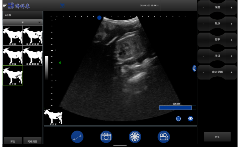
B-mode (brightness mode) ultrasound remains the most common and effective modality for fetal sexing. It provides a two-dimensional, real-time grayscale image of the fetus inside the uterus. Operators move a linear or convex rectal probe inside the cow to scan the uterine horns and locate the fetus.
Once located, they carefully examine the perineal and abdominal area of the fetus for the genital tubercle’s position. Patience and practice are essential—especially in early gestation when the fetus is small and mobile.
This non-invasive, repeatable, and relatively low-cost technique has become a standard tool in progressive cattle operations. It’s especially favored for:
-
Speed: A trained operator can sex a fetus in under two minutes.
-
Safety: No drugs or invasive tools are used.
-
Real-time feedback: Images can be captured and stored for later review or validation.
Practical Considerations in the Field
-
Restraint and Positioning
The cow must be properly restrained in a chute or crush. Movement during the scan can obscure the fetal view and reduce accuracy. -
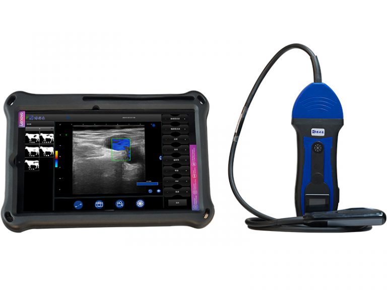
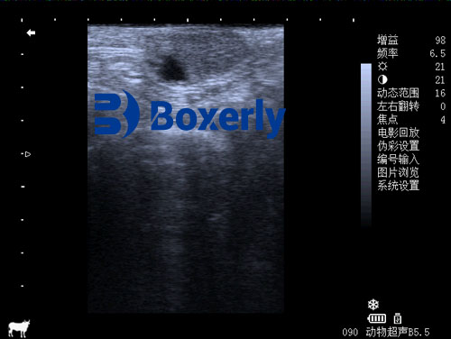
Ultrasound Equipment Quality
A high-resolution veterinary ultrasound device with a rectal probe improves visualization, especially under field conditions. For instance, B-mode machines like those used in North American and European farms allow precise localization of the genital tubercle with little guesswork. -
Fetal Orientation
Sometimes, the fetus may be facing away or curled in a way that obscures genital structures. In such cases, scanning may need to be rescheduled within the ideal gestation window. -
Operator Skill
Reproductive ultrasound is highly operator-dependent. Training and experience are critical. Many farms send their technicians to specialized training programs in Australia, Canada, or the U.S. to master fetal sexing techniques.
What Can Go Wrong?
Even with the best equipment and a skilled operator, fetal sex determination isn’t infallible. Potential issues include:
-
Misidentification of genital structures: A misread position of the genital tubercle may lead to an incorrect call.
-
Late or early scanning: Scanning before day 55 or after day 90 increases error rates.
-
Cow movement: Fetal orientation may shift due to movement or contractions.
-
Equipment limitations: Older or low-resolution ultrasound units can fail to provide a clear image.
Farmers and vets understand these challenges and often re-confirm the diagnosis on a second scan or wait until birth for absolute certainty. Nevertheless, even a 90% confidence level has proven beneficial for herd-level decision-making.
International Perspectives and Adoption
In countries like the U.S., large-scale dairy farms use fetal sexing to identify female calves early and selectively cull or repurpose pregnancies carrying male calves. In beef operations, this information helps optimize feed costs and marketing schedules.
In Australia, fetal sexing has been widely adopted on ranches using artificial insemination or embryo transfer. Some veterinarians even offer sexing services as part of routine pregnancy checks, supported by portable, rugged ultrasound machines ideal for field use.
In Europe, especially in Germany and the Netherlands, sexed semen and fetal ultrasound are paired to ensure high accuracy in heifer production.
These global practices highlight the role of ultrasound not just in diagnostics but as a strategic breeding and marketing tool.

The Economic Impact of Knowing Fetal Sex
The cost of a routine pregnancy ultrasound scan is relatively low. Adding a fetal sexing examination typically adds little to the cost but significantly boosts value.
Producers can make early sales decisions, decide whether to induce abortion (in extreme cases), or adjust nutrition and management for dams carrying more valuable calves.
In embryo transfer programs, knowing the fetal sex supports better herd planning. For instance, if producing high-value replacement heifers, only pregnancies with confirmed female fetuses may be kept.
Studies in Canada and the U.S. suggest that farms utilizing fetal sexing with ultrasound report better reproductive efficiency, lower heifer rearing costs, and faster herd genetic gains.
Future Developments
Ultrasound technology continues to evolve. Modern portable ultrasound machines with higher resolution, wireless capabilities, and smart image processing make fetal sexing even easier and more accurate.
Training has also become more accessible, with online modules and hands-on workshops now available globally. AI-assisted imaging and automated fetal sex prediction tools are being researched, which could further reduce operator dependence in the near future.
As demand for sexed pregnancies rises—especially in precision breeding and conservation herds—ultrasound will remain a vital tool in the toolbox of modern livestock producers.
Conclusion
Determining the sex of a cow’s fetus by ultrasound offers tangible benefits to cattle producers around the world. It enables early management decisions, improves economic returns, and supports herd genetic planning.
With proper equipment, training, and timing, B-mode ultrasound allows veterinarians and producers to identify fetal sex with over 90% accuracy during the 60–90 day gestation window. This simple, non-invasive method has become standard practice in many progressive operations and continues to grow in popularity thanks to its proven value and growing accessibility.
As the livestock industry leans more toward data-driven and precision management, tools like ultrasonography will continue to play a central role—not just in detecting pregnancy, but in shaping the future of breeding and herd development.
References
-
Fricke, P. M. (2019). “Ultrasound-based fetal sexing in dairy cattle.” Journal of Dairy Science, 102(6), 5671–5682. https://doi.org/10.3168/jds.2018-15800
-
Curran, S., Pierson, R. A., & Ginther, O. J. (1989). “Ultrasonographic appearance of the bovine conceptus from days 20 through 60.” Journal of the American Veterinary Medical Association, 195(4), 466–470. https://pubmed.ncbi.nlm.nih.gov/2676914/
-
Australian Cattle Veterinarians (2024). “Reproductive Ultrasound Guidelines.” https://www.cattlevets.com.au/ultrasound
-
University of Wisconsin Extension. (2023). “Using Ultrasound for Fetal Sex Determination in Cattle.” https://fyi.extension.wisc.edu/dairy/ultrasound-fetal-sexing