How Farms Detect Pig Ovarian Cysts With Ultrasound
For many pig farmers, maintaining the reproductive health of their sows is essential to the economic success of their operations. But not every fertility issue is visible on the surface—some remain hidden within the reproductive tract, silently disrupting breeding plans and lowering farrowing rates. One of the most common yet elusive reproductive disorders in sows is the ovarian cyst. These cysts can delay or even completely block ovulation, leaving a seemingly healthy sow unproductive. That’s where ultrasound comes in.

Over the past decade, farms around the world have turned to ultrasound as a go-to diagnostic tool—not just for pregnancy checks but for investigating deeper issues like ovarian cysts. This isn’t just a high-tech add-on; it’s becoming a routine part of herd reproductive management in many modern pig farms, from Iowa to Denmark.
Understanding Ovarian Cysts in Pigs
Ovarian cysts are fluid-filled structures that develop on a sow’s ovaries. They usually fall into two categories: follicular cysts (which prevent ovulation) and luteal cysts (which can interfere with the hormonal cycle). Both types affect fertility, but their causes can vary—from nutritional imbalances and stress to hormonal disturbances or incorrect estrus synchronization protocols.
What makes ovarian cysts especially frustrating for farmers is that affected sows may still show signs of heat or may seem outwardly normal. Without internal examination, these cysts can go completely unnoticed until breeding repeatedly fails.
Why Ultrasound Is a Game-Changer
Before ultrasound became widely used, diagnosing ovarian cysts involved a lot of guesswork, often relying on hormonal treatments to “see what happens” or waiting for multiple failed breedings before taking action. This not only wasted time and money but also led to unnecessary culling of potentially valuable animals.
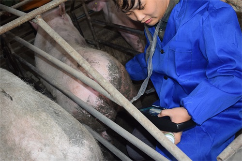
Today, farms are using real-time B-mode ultrasound to look directly at the ovaries. A skilled technician can quickly scan through the abdominal wall or rectally (depending on the sow’s condition and scanner type) and detect enlarged fluid-filled structures that indicate cysts. In some cases, Doppler ultrasound is used to analyze blood flow and differentiate between cyst types.
Ultrasound is:
-
Non-invasive and stress-free
-
Fast—a typical scan takes less than five minutes
-
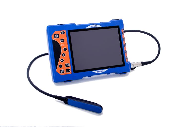
Portable, with handheld devices now common on farms
-
Cost-effective when used regularly across a herd
What an Ultrasound Technician Looks For
When scanning for ovarian cysts, several signs stand out:
-
Follicular cysts appear as round, anechoic (black) fluid-filled structures larger than normal follicles—usually more than 2.5 cm in diameter.
-
Luteal cysts may appear slightly more echogenic (grayish) with thicker walls and sometimes vascularized (visible blood flow).
-
Multiple cysts often cluster, giving the ovary a “bunch of grapes” appearance on screen.
-
Lack of corpus luteum in what should be a post-ovulation period may also signal cystic ovaries.
On some farms, technicians conduct follow-up scans days or weeks later to monitor changes and assess the effectiveness of treatment protocols.
The Practical Benefits for Farmers
Detecting ovarian cysts with ultrasound isn’t just about knowing what’s wrong—it’s about knowing what to do next. Based on the scan results, farmers and vets can decide whether to:
-
Apply hormonal treatment (e.g., GnRH or PGF2α)
-
Cull the sow if prognosis is poor
-
Adjust feeding or management to reduce stress-related triggers
-
Modify estrus synchronization protocols across the herd
This level of precision reduces unnecessary treatments, improves reproductive efficiency, and helps keep more productive sows in the herd. Some producers report a 10–15% improvement in breeding success rates simply by adding regular ovary scanning to their herd health plans.
Ultrasound Integration on Modern Pig Farms
Ultrasound use isn’t limited to large commercial farms anymore. With handheld, battery-powered devices becoming cheaper and more user-friendly, even small family farms are integrating ultrasound into their breeding routines.
In countries like the Netherlands, Canada, and Germany, it’s now common to see producers conducting their own scans after a few hours of training. They typically check sows:
-
Before artificial insemination, to confirm the presence of mature follicles
-
After repeated breeding failure, to rule out cysts or confirm anestrus
-
During mid-pregnancy, to monitor gestation and fetal health
What’s more, some producers scan boars as part of fertility assessments, especially when poor conception rates are suspected. While boars don’t get ovarian cysts (obviously), ultrasound can detect abnormalities in the testes or accessory glands that may affect fertility.
Limitations and Considerations
While ultrasound is powerful, it’s not infallible. A few key things to keep in mind:
-
Operator experience matters: Misreading a structure on-screen can lead to misdiagnosis. Training and practice are critical.
-
Some cysts are transient: Not all detected cysts require treatment. Some resolve on their own.
-
Scan timing matters: Ovarian structures change quickly across the cycle. Scanning at the wrong time can confuse diagnosis.
-
Not a stand-alone solution: Ultrasound should be part of a broader reproductive management plan, including good record-keeping and nutrition.
Despite these challenges, most producers find that the benefits of early, accurate diagnosis far outweigh the learning curve.
Global Perspectives on Swine Ultrasound Use
Across Europe and North America, the integration of ultrasound into swine health programs has been well documented. In Denmark, a country known for its high-efficiency pig farming, routine ovary scanning has been part of national herd management protocols for years. Vets there use it not just for diagnosis but also for herd fertility benchmarking.
In the U.S., especially in high-production states like North Carolina and Minnesota, vets are increasingly recommending ovarian scanning post-weaning to catch early signs of cyst development. Producers find this especially helpful for identifying “silent non-breeders”—sows that appear healthy but quietly fail to cycle.
Even in developing countries, where veterinary resources may be more limited, portable ultrasound devices are being introduced through government or university extension programs. These tools are helping smallholder farmers improve sow productivity, reduce culling rates, and increase litter sizes over time.
The Future of Reproductive Monitoring
As ultrasound technology continues to evolve, we may soon see even more precise diagnostic capabilities. Some manufacturers are developing AI-assisted image recognition systems that automatically flag cysts or irregular follicle patterns. Combined with farm data management apps, this could help producers track ovarian health trends across the herd.
There’s also growing interest in tele-ultrasound, where on-farm scans are remotely interpreted by a vet via smartphone or cloud connection. This could be a game-changer for smaller farms without direct vet access.
Summary
On today’s pig farms, staying ahead means understanding more than just when a sow is pregnant—it’s about knowing why she’s not. Ultrasound offers that deeper layer of insight, especially when it comes to detecting ovarian cysts.
By incorporating regular ovary scanning into their management routines, producers can catch reproductive issues early, apply targeted treatments, and avoid unnecessary losses. Whether it’s through a vet visit or a handheld scanner on the back of a pickup truck, this technology is helping pig farms big and small improve fertility, efficiency, and profit.