How Farms Detect Horse Tendon Injuries with Ultrasound
For horse owners, trainers, and farm vets, tendon injuries are among the most dreaded diagnoses. They not only interrupt a horse’s training or competition season but often require months of rehabilitation and can even end a promising career. That’s why early detection and accurate diagnosis are critical—and this is where ultrasound plays a game-changing role on farms around the world.
Across equine facilities, from elite racing stables in Kentucky to small family farms in rural Europe, ultrasound has become a go-to tool for monitoring tendon health. It’s non-invasive, safe, and offers detailed insights that can’t be picked up through palpation alone. Let’s take a closer look at how horse farms use ultrasound to detect tendon injuries and why it’s now considered essential in modern equine care.
Why Tendon Health Matters in Horses
Tendons are crucial for locomotion. In horses, the superficial digital flexor tendon (SDFT) and deep digital flexor tendon (DDFT) are especially prone to injury, particularly in athletic animals. When these tendons get overworked or overstretched—whether during racing, jumping, or simply uneven terrain—they can suffer micro-tears or more severe damage.
Once injured, tendons heal slowly and are prone to re-injury. That’s because tendinous tissue heals with scar formation rather than regenerating the original elastic fibers. Scar tissue is less elastic and more vulnerable under strain. So, the earlier a problem is found, the better the long-term outcome.
Palpation vs. Ultrasound: What Farmers and Vets Have Learned
Many farm owners still rely on palpation as a first check. And it’s true—palpating the tendon line can reveal heat, swelling, or a soft spot that suggests injury. But here’s the problem: by the time those symptoms are visible or feelable, the damage is already done.
That’s why experienced farm vets now turn to ultrasound early, especially when a horse is suddenly off, sensitive, or shows even minor changes in gait. Ultrasound lets them look beneath the skin—literally.
Using a linear ultrasound probe with frequencies between 7.5 to 15 MHz, the vet can visualize the internal structure of the tendon, spotting disruptions in fiber alignment, fluid accumulation, or inflammation. They can even detect minor lesions that would otherwise go unnoticed.
What the Ultrasound Actually Shows
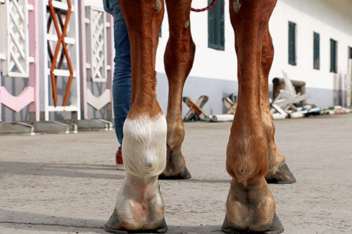
When a vet performs an ultrasound on a horse’s leg, they usually scan the palmar or plantar area (back side of the leg), starting from just below the carpus (knee) or hock and moving down toward the fetlock.
A healthy tendon shows up as a series of parallel echogenic lines—think of them as tidy, white stripes. These represent aligned collagen fibers. When there’s an injury, those lines appear disrupted. You might see dark (hypoechoic) areas, which indicate fluid or fiber rupture, or a general swelling of the tendon.
Ultrasound also allows for measuring:
-
Cross-sectional area (CSA) of the tendon
-
Echogenicity (brightness, linked to inflammation or fiber integrity)
-
Fiber alignment
-
Peritendinous fluid (fluid surrounding the tendon)
These measurements can be recorded over time, helping track healing or identify re-injury risk.
Common Scenarios Where Ultrasound Saves the Day
Here’s how farms typically use ultrasound for tendon management:
1. Routine Baseline Scans
Top-level performance stables often conduct pre-season or routine scans on their horses. These “baseline” images become a comparison point for any future scans, making it easier to catch subtle changes.
2. Early Lameness Investigation
If a horse goes slightly lame, even without visible swelling, ultrasound can determine whether the tendon is the source. Many tendon injuries start subtly, with micro-damage before the tendon actually tears.
3. Post-Injury Monitoring
Once a tendon injury has been diagnosed, ultrasound helps guide the rehab process. Vets will schedule follow-up scans every 4–6 weeks to assess healing progress and adjust the recovery program accordingly.
4. Pre-Purchase Exams
In high-value sales, ultrasound scans of the tendons are often included in veterinary pre-purchase exams. They help ensure there are no pre-existing tendon weaknesses that could spell future trouble.
Real Talk from the Field: What Foreign Vets Are Saying
Across Europe, Australia, and North America, equine practitioners agree—ultrasound has become a diagnostic pillar in equine lameness evaluation. Here’s a look at a few observations from vets and trainers abroad:
Dr. Katie Thomlinson (UK): “We use ultrasound almost every day now in equine practice. The ability to detect early tendon strain before it becomes a tear has saved careers—literally.”
Mark Pelham, trainer (New Zealand): “We lost a horse a few years ago to a tendon rupture that might’ve been avoided if we’d caught it sooner. Now, we scan every six weeks during competition season. It’s worth the peace of mind.”
Dr. Jeremy Roberts (USA): “What ultrasound gives us isn’t just data—it’s confidence. When we clear a horse to return to work, we know it’s based on solid evidence, not guesswork.”
Pros and Limitations of Ultrasound in Tendon Injury Detection
👍 Pros:
-
Non-invasive and repeatable
-
Detects both acute and chronic changes
-
Can guide both diagnosis and treatment
-
Portable systems allow stall-side exams
-
Helps predict re-injury risk during rehab
👎 Limitations:
-
Requires training to interpret images correctly
-
Can miss deeper soft tissue issues without experience
-
Not ideal for detecting very early biochemical changes (e.g., cellular-level inflammation)
Despite these minor limitations, when combined with clinical exams and experienced hands, ultrasound remains the best first-line imaging tool for equine tendon assessment on the farm.
Tips for Farm Owners: Making the Most of Equine Ultrasound
If you run a farm or stable, here are a few practical ways to incorporate ultrasound into your tendon injury prevention strategy:
-
Partner with a vet experienced in equine ultrasound
-
Create tendon “profiles” for all working horses
-
Don’t wait for lameness—use ultrasound at the first sign of discomfort
-
Keep detailed records of scans for long-term comparison
-
Follow vet advice closely during recovery, using ultrasound to guide rest and rehabilitation timelines
A Glimpse into the Future
Advanced ultrasound techniques like elastography and Doppler imaging are slowly making their way into equine sports medicine. Elastography measures tendon stiffness, offering earlier detection of subtle changes in tendon properties before any visible damage occurs. Color Doppler can highlight blood flow patterns in healing tissue, giving vets an edge in assessing inflammation and tissue repair.
While these are still more common in equine hospitals than on small farms, portable ultrasound technology is catching up fast. That means even everyday farm owners might soon have access to hospital-grade insights—right in the barn.
Final Thoughts
Tendon injuries may be part of horse life, but that doesn’t mean they have to end a career. With today’s ultrasound technology, farm vets and horse owners can work together to catch problems early, monitor healing with precision, and make smarter decisions about training and rest. The ability to look inside the leg, assess real-time tendon health, and respond quickly is a game changer—especially when the health and future of a beloved horse are on the line.