How Does Veterinary Color Doppler Ultrasound Work?
Ultrasound technology has transformed the way veterinarians diagnose and monitor animal health, especially in the livestock and farm animal industries. Among the various ultrasound techniques available, Color Doppler ultrasound stands out as a powerful, non-invasive tool that provides real-time insight into blood flow dynamics. For veterinarians and animal breeders alike, understanding how Color Doppler ultrasound works and what benefits it brings can significantly improve reproductive management, early disease detection, and overall herd productivity.

In this comprehensive guide, we’ll explore the science behind veterinary Color Doppler ultrasound, its practical applications in large animal care, and how it can enhance efficiency and decision-making in your veterinary practice or farm operation.
What Is Color Doppler Ultrasound?
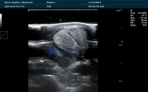
Color Doppler ultrasound is a specialized imaging technique that uses standard ultrasound technology combined with the Doppler effect to visualize and measure blood flow within the body. Unlike traditional B-mode ultrasound—which creates grayscale images based on tissue density—Color Doppler adds color overlays that indicate the direction and velocity of blood flow within vessels and organs.
This enhanced capability enables veterinarians to observe and assess circulation patterns, detect abnormalities in blood flow, and evaluate organ perfusion in real time.

The Science Behind Doppler Ultrasound
To understand how Color Doppler ultrasound works, let’s first look at the Doppler effect. This is the same principle that causes the pitch of a siren to change as an ambulance drives past.
In medical ultrasound, sound waves are transmitted into the body via a transducer. When these waves encounter moving red blood cells within vessels, they reflect back at different frequencies depending on the speed and direction of the blood flow:
-
Flow toward the transducer increases the frequency of the reflected sound waves.
-
Flow away from the transducer decreases the frequency.
The ultrasound machine calculates these frequency shifts and translates them into visual data. The result is a real-time color map superimposed on the grayscale anatomical image, with:
-
Red typically indicating flow toward the transducer.
-
Blue indicating flow away from the transducer.
-
Variations in brightness or hue corresponding to the velocity of the flow.
This data enables precise assessment of vascular structures, blood circulation, and even organ function.
Types of Doppler Ultrasound Modes
Veterinary Color Doppler systems often include multiple Doppler modes, each offering different levels of detail:
-
Color Flow Doppler
This mode displays blood flow direction and velocity as a color map. It’s ideal for quickly identifying vascular structures and assessing general flow patterns. -
Power Doppler
This mode detects the strength (amplitude) of the Doppler signal, making it more sensitive to low-flow states. It’s especially useful in small or deep vessels where standard color Doppler may be less effective. -
Spectral Doppler (Pulsed-Wave and Continuous-Wave)
This provides a graphical representation of flow velocity over time. Pulsed-wave Doppler is used for measuring flow in a specific location, while continuous-wave Doppler is better for detecting high-velocity flows across heart valves or large vessels. -
Triplex Imaging
A combination of B-mode, Color Doppler, and Spectral Doppler imaging displayed simultaneously. This integrated approach improves diagnostic confidence by correlating anatomical and hemodynamic data in real time.
How It Works in Veterinary Practice
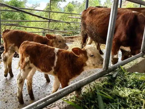
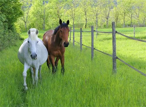
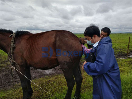
Color Doppler ultrasound is applicable to a wide range of veterinary procedures, particularly in large animals such as cattle, horses, sheep, goats, pigs, and even exotic farm animals. Let’s examine some of its most impactful uses in the field:
1. Reproductive Monitoring and Fertility Evaluation
In livestock production, reproduction efficiency directly impacts profitability. Color Doppler ultrasound can significantly improve reproductive outcomes through:
-
Follicular and luteal blood flow assessment: The vascularity of the corpus luteum or ovarian follicles is a key indicator of reproductive status and ovulation potential.
-
Early pregnancy confirmation: Increased blood flow to the uterus can indicate early implantation or placental development.
-
Uterine health evaluation: Doppler imaging helps detect uterine inflammation (endometritis) or infections by analyzing vascular changes.
-
Male fertility screening: Blood flow to the testes and spermatic cord is important for normal spermatogenesis; Color Doppler helps identify testicular torsion, varicocele, or hypoplasia.
2. Cardiovascular Assessment
Heart disease is not just a concern in companion animals. In working horses and high-value livestock, cardiac evaluation is vital for long-term health and performance. Doppler ultrasound enables:
-
Detection of congenital heart defects
-
Assessment of valve function and regurgitation
-
Measurement of cardiac output and blood velocity
-
Monitoring post-operative or post-treatment recovery
Spectral Doppler is particularly useful in equine cardiology, where evaluating pulmonary artery pressures and heart valve integrity is critical.
3. Liver, Kidney, and Spleen Evaluation
Color Doppler ultrasound allows visualization of vascular perfusion in vital organs:
-
Liver: Detects portal hypertension, thrombosis, or hepatopathies in cattle and sheep.
-
Kidneys: Measures renal arterial flow to detect obstruction, infarction, or chronic nephritis.
-
Spleen: Identifies splenic torsion or abnormal congestion in pigs or ruminants.
Changes in blood flow patterns often precede changes in organ size or structure, making Color Doppler valuable for early intervention.
4. Musculoskeletal Injuries and Tendon Healing
In working and racing animals like horses and camels, muscle and tendon injuries are common. Doppler ultrasound is used to:
-
Evaluate blood supply to injured tendons
-
Monitor healing progress and angiogenesis
-
Determine the appropriate time for return to work
This can dramatically reduce the risk of re-injury and optimize recovery protocols.
5. Oncology and Tumor Evaluation
One of the most promising applications of Color Doppler is in oncology:
-
Vascularity analysis of tumors: Malignant masses often have increased and chaotic blood flow.
-
Differentiation between benign and malignant lesions
-
Guidance for fine-needle aspiration or biopsy
In large animals, early detection of tumors can be lifesaving and cost-effective, especially in breeding stock.
Advantages of Using Color Doppler in Livestock
Investing in veterinary Color Doppler ultrasound offers several benefits for farm operations and animal health monitoring:
| Advantage | Explanation |
|---|---|
| Early Diagnosis | Blood flow changes can indicate disease before structural damage appears. |
| Non-Invasive | No sedation, radiation, or surgical exposure required. |
| Real-Time Feedback | Immediate visualization of physiological processes. |
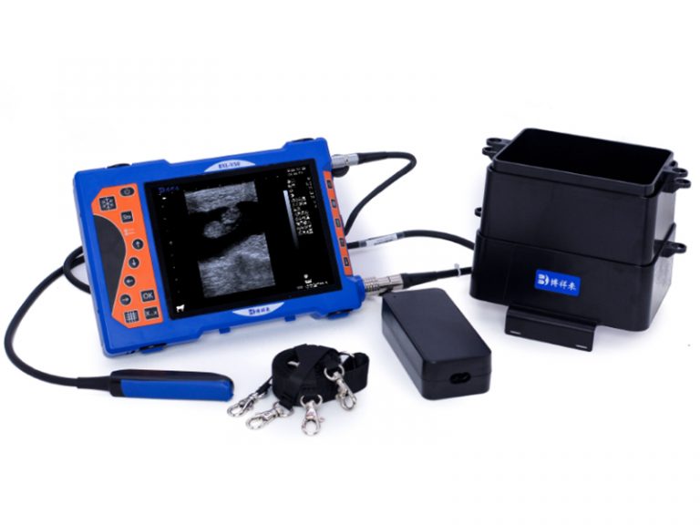
| Portable and Field-Ready | Many systems are rugged and battery-powered for on-site use. |
| Better Reproductive Outcomes | More precise timing and monitoring of breeding efforts. |
| Cost Savings | Reduces the need for exploratory surgery or unnecessary treatments. |
Choosing the Right Doppler Ultrasound for Veterinary Use
Not all Color Doppler ultrasound machines are equally suited for livestock. When selecting a system, consider:
-
Probe types: Convex or micro-convex for abdominal scans; linear for superficial vessels or tendon imaging.
-
Frequency range: Lower frequencies (2–5 MHz) for deep structures in cattle and horses; higher frequencies (7.5–12 MHz) for small ruminants or reproductive organs.
-
Battery life and portability: Essential for mobile farm visits.
-
Ease of use: Intuitive interfaces and automated measurement tools save time.
-
Image quality and Doppler sensitivity: High frame rates and color sensitivity ensure accuracy.
-
Brand and support: Look for brands like BXL that specialize in veterinary applications and offer solid training and technical support.
Practical Tips for Effective Use
To get the most from your Color Doppler ultrasound system:
-
Ensure good contact with the skin using appropriate gel or coupling agents.
-
Minimize motion artifacts by calming the animal or using holding chutes.
-
Adjust color gain and wall filters to optimize flow visualization.
-
Scan at a proper angle (ideally < 60°) to avoid Doppler angle distortion.
-
Record and compare results over time to monitor treatment or reproductive progress.
Training is key—consider investing in ultrasound workshops or online courses focused on veterinary Doppler techniques.
Future Developments in Veterinary Doppler Ultrasound
The field of veterinary imaging continues to evolve rapidly. Emerging advancements include:
-
AI-assisted flow analysis: Automated detection of abnormal patterns.
-
Wireless probes and tablet-based systems: Enhanced mobility and telemedicine support.
-
Elastography integration: Assessment of tissue stiffness alongside vascularity.
-
3D/4D Doppler imaging: Real-time, volumetric views of complex blood flow.
As these technologies become more accessible, Color Doppler ultrasound will become an indispensable tool for any livestock veterinarian or breeder aiming for top-tier animal health management.
Conclusion
Veterinary Color Doppler ultrasound offers a powerful, real-time window into the circulatory system of animals. From improving fertility rates to detecting life-threatening conditions early, it enables better, faster, and more accurate decision-making in livestock care.
Whether you’re managing a herd of dairy cows, breeding performance horses, or treating farm animals in the field, integrating Color Doppler into your veterinary toolkit can raise the standard of care and contribute directly to the health, productivity, and profitability of your operation.
For veterinarians and animal producers striving to stay ahead in today’s competitive market, Color Doppler ultrasound isn’t just an option—it’s a necessity.