
Cow Gestational Sac Ultrasound: How to Detect Early Pregnancy in Cattle with Veterinary Ultrasound

Cow Gestational Sac Ultrasound: How to Detect Early Pregnancy in Cattle with Veterinary Ultrasound
Introduction: The Importance of Gestational Sac Ultrasound in Cattle
Early pregnancy diagnosis is an essential part of modern cattle reproductive management. For dairy farms, beef cattle operations, breeding centers, and veterinary clinics, identifying pregnant cows as early as reliably possible helps improve breeding efficiency and reduce unnecessary reproductive delays.
One of the important findings during early pregnancy examination is the gestational sac, also known as the embryonic vesicle.
Cow gestational sac ultrasound allows veterinarians to examine the uterus and identify pregnancy-related structures using real-time ultrasound imaging. With an appropriate transrectal ultrasound scanner, veterinarians can observe the developing gestational sac, evaluate its appearance, and monitor subsequent embryonic development.
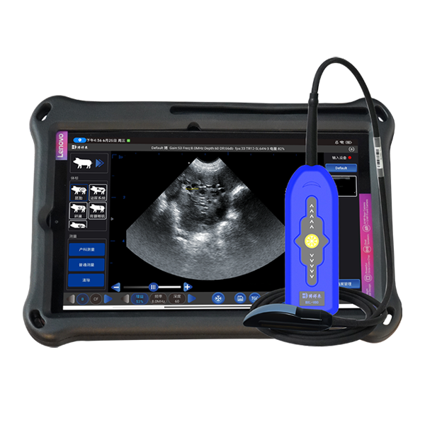
Compared with traditional manual examination, ultrasound provides direct visual information about the reproductive tract.
For cattle veterinarians and breeding specialists, a high-quality veterinary ultrasound machine is an important tool for early pregnancy diagnosis, uterine examination, and reproductive monitoring.
In this article, we explain how gestational sac ultrasound works in cattle, when the gestational sac can be detected, and what features to consider when choosing a professional cattle pregnancy ultrasound scanner.
1. What Is a Gestational Sac in Cattle?
The gestational sac is an early pregnancy-related structure that develops within the uterus following successful fertilization and embryonic development.
In cattle, the early conceptus develops within the uterine environment. As development progresses, the embryonic membranes form fluid-containing structures that can be identified through ultrasound examination.
On a B-mode ultrasound image, the early gestational sac generally appears as a fluid-filled area with relatively dark, anechoic contents surrounded by brighter tissue interfaces.
As pregnancy progresses, additional structures become visible, including:
The developing embryonic vesicle.
The embryo proper.
Embryonic membranes.
The amniotic region.
The developing fetal structures.
The appearance of these structures changes with gestational age.
Identifying the gestational sac is an important step in early bovine pregnancy examination. However, the presence of a fluid-filled structure alone should not automatically be interpreted as definitive pregnancy confirmation. Veterinarians should evaluate its location, appearance, development, and associated embryonic findings.
2. How Does Cow Gestational Sac Ultrasound Work?
<IMAGE_PLACEHOLDER>
Veterinary ultrasound imaging uses high-frequency sound waves to produce real-time images of internal reproductive structures.
For cattle pregnancy diagnosis, veterinarians commonly use a transrectal linear probe to examine the uterus through the rectal wall.
The ultrasound probe sends sound waves into the reproductive tract and receives the returning echoes.
The ultrasound system processes these signals and displays a grayscale image on the screen.