Ultrasound for Sheep Pregnancy: A Practical Guide to Pregnancy Scanning and Veterinary Ultrasound Equipment

Ultrasound for Sheep Pregnancy: A Practical Guide to Pregnancy Scanning and Veterinary Ultrasound Equipment
Ultrasound for sheep pregnancy has become an important tool for modern sheep farming, helping farmers and veterinarians assess reproductive status and improve flock management. With a suitable veterinary ultrasound scanner, trained operators can examine pregnant ewes, identify pregnancy at an appropriate stage, and obtain useful information to support breeding and lambing preparation.
For sheep farmers, livestock breeding organizations, and veterinary professionals, choosing the right sheep pregnancy ultrasound machine is an important investment. Image quality, probe selection, portability, battery performance, ease of operation, and technical support should all be considered before purchasing equipment.
Zhengzhou Boxianglai Electronic Sci-Tech Co., Ltd., operating under the BOXERLY brand, specializes in veterinary ultrasound equipment for livestock applications. With in-house manufacturing capabilities and product development experience, BOXERLY provides portable ultrasound solutions for international customers seeking practical equipment for animal reproductive examinations.

This guide explains how ultrasound for sheep pregnancy works, when to scan ewes, what to consider when selecting a sheep ultrasound scanner, and how BOXERLY can support your equipment requirements.
1. Why Is Ultrasound for Sheep Pregnancy Important?
Efficient reproductive management is essential for sheep farms of different sizes. Without appropriate pregnancy diagnosis, farmers may find it difficult to organize breeding records, manage feeding programs, and prepare for lambing.
A suitable sheep pregnancy scanner allows trained operators to assess pregnancy status through ultrasound imaging. The results can support more systematic decisions throughout the breeding cycle.
Pregnancy Diagnosis
Ultrasound examination can help identify pregnant ewes at an appropriate stage of gestation. Early confirmation allows farmers to review breeding records and organize follow-up examinations when necessary.
Flock Management
Grouping ewes according to reproductive status can help farms plan daily management activities. Ultrasound findings can contribute to identifying animals that require further examination or different management arrangements.
Feeding and Nutritional Planning
Pregnancy status is one factor considered when planning ewe nutrition. Farmers can use examination results together with veterinary advice, body condition, and production requirements to develop appropriate feeding strategies.
Preparation for Lambing
Reliable breeding records and pregnancy assessments can help farmers estimate lambing periods and organize labor, housing, and other resources.
Reproductive Monitoring
For farms with structured breeding programs, ultrasound examinations can contribute to routine reproductive monitoring and more complete animal records.
The benefits depend on correct scanning techniques, suitable equipment, and appropriate interpretation of the images. Ultrasound findings should be assessed by a trained veterinarian or qualified operator.
2. When Can You See Sheep Pregnancy with Ultrasound?
One of the most common questions from sheep farmers is: How early can an ultrasound scanner detect pregnancy in sheep?
The answer depends on the examination method, probe, image quality, ewe condition, and operator experience.
The following timeline provides general guidance.
Around 20–25 days after mating: Very early pregnancy structures may be detectable under suitable conditions, but examinations at this stage can be challenging. A negative or uncertain result may require a follow-up scan.
Around 25–35 days: Pregnancy structures and the developing embryo are generally easier to identify as gestation progresses. Detection of the embryo and heartbeat depends on the actual gestational age and examination conditions.
Around 35–50 days: The developing fetus becomes more recognizable, and major anatomical structures may be visible in suitable ultrasound views.
Around 50–70 days: The fetus is larger, making some structures easier to identify. However, seeing the entire fetus in one image is not always possible because of fetal size, position, and the scanner’s field of view.
For farmers who want to demonstrate recognizable fetal anatomy, scanning at approximately 40–60 days of gestation can be a practical starting point, although results vary.
The appropriate examination time should be selected according to the purpose of the scan. Early pregnancy diagnosis and detailed fetal visualization are different objectives and may require different scanning approaches.
3. What Can a Sheep Pregnancy Ultrasound Scanner Show?
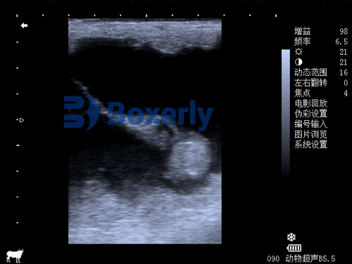
A veterinary ultrasound scanner produces images from returning sound waves, allowing trained operators to examine relevant structures inside the animal.