Early Embryonic Pregnancy Diagnosis in Cattle: How Ultrasound Helps Detect Pregnancy Earlier and Improve Reproductive Management

Early Embryonic Pregnancy Diagnosis in Cattle: How Ultrasound Helps Detect Pregnancy Earlier and Improve Reproductive Management
1. Why Is Early Embryonic Pregnancy Diagnosis Important in Cattle?
Early pregnancy diagnosis is an important part of modern cattle reproductive management.
For dairy farms, beef cattle operations, and professional breeding centers, identifying pregnant and non-pregnant cows at an appropriate time can help improve breeding efficiency and reduce unnecessary waiting periods.
When pregnancy status remains unknown for too long, farm managers may miss opportunities to make timely reproductive management decisions.
This is particularly important in large cattle operations where hundreds or thousands of breeding animals may need regular reproductive monitoring.
Early embryonic pregnancy diagnosis in cattle focuses on identifying early pregnancy-related structures and evaluating embryonic development before the pregnancy progresses to a more advanced stage.
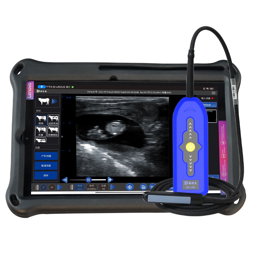
Veterinary ultrasound has become an important diagnostic tool for this purpose.
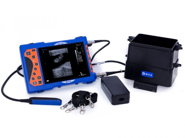
Using a suitable veterinary ultrasound scanner equipped with a professional cattle rectal linear probe, veterinarians can examine:
– Early pregnancy-related uterine changes.
– The gestational sac.
– The developing embryo.
– Embryonic cardiac activity at an appropriate stage.
– The corpus luteum.
– Ovarian structures.
– Uterine conditions.
Unlike pregnancy tests that primarily provide indirect information, ultrasound allows veterinarians to visualize reproductive structures in real time.
This makes it particularly valuable for cattle breeding programs that require detailed reproductive examinations.
2. When Can Early Embryonic Pregnancy Be Detected in Cattle?
One of the most common questions from cattle farmers and veterinarians is:
How early can you detect pregnancy in a cow using ultrasound?
The answer depends on several factors, including gestational age, ultrasound equipment quality, probe frequency, examination technique, and operator experience.
During early pregnancy, the embryo is very small, so identifying pregnancy-related structures requires suitable imaging equipment and professional scanning skills.
The following table provides general reference information.
Days after insemination Possible ultrasound findings
18–21 days Early reproductive changes may occur, but routine ultrasound pregnancy confirmation is unreliable
22–25 days Some early uterine changes may be observed, but embryo identification is challenging
25–28 days Gestational structures may become detectable in suitable cases
28–30 days Gestational sac and embryo may be identifiable by experienced operators
30–35 days Early pregnancy confirmation becomes more practical
35–45 days Embryonic structures and cardiac activity can generally be evaluated more reliably
After 45 days Further embryonic and early fetal development assessment becomes possible
These are approximate clinical reference windows, not guaranteed detection dates. Actual results depend on the animal, equipment, examination technique, and operator experience.
For routine pregnancy confirmation, many veterinarians prefer scanning around 28–35 days after insemination or later, depending on their diagnostic protocol.
An examination performed too early may produce an uncertain result. When pregnancy cannot be confirmed, a repeat scan is recommended.
3. How Does Ultrasound Detect Early Embryonic Pregnancy in Cattle?
Veterinary ultrasound uses high-frequency sound waves to produce images of internal reproductive structures.
For early cattle pregnancy examinations, transrectal ultrasound is widely used because it allows convenient access to the uterus and ovaries.
A suitable rectal linear probe provides real-time imaging that helps veterinarians examine changes associated with early pregnancy.
There are several important structures to identify.
3.1 Gestational Sac
The gestational sac is one of the earliest recognizable pregnancy-related structures.
On ultrasound, it appears as a fluid-filled area within the uterus.
Its location, appearance, and development help veterinarians evaluate early pregnancy.
3.2 Developing Embryo
As pregnancy progresses, the embryo becomes visible within the gestational sac.
At this stage, a high-quality ultrasound image is important because the embryo is still relatively small.
Veterinarians assess its appearance together with other pregnancy-related findings.
3.3 Embryonic Heartbeat
At an appropriate developmental stage, embryonic cardiac activity can be observed using real-time B-mode ultrasound.