
Colour Doppler Pregnancy Diagnosis in Cows: Advanced Ultrasound Technology for Early Cattle Pregnancy Detection

Colour Doppler Pregnancy Diagnosis in Cows: Advanced Ultrasound Technology for Early Cattle Pregnancy Detection
Introduction: A New Approach to Pregnancy Diagnosis in Cows
Efficient reproductive management is essential for profitable dairy and beef cattle production. Every unsuccessful breeding cycle can increase production costs, extend calving intervals, and reduce overall herd efficiency.
For veterinarians and cattle breeders, identifying pregnancy as early as reasonably possible is therefore an important part of herd management.
Traditional B-mode ultrasound provides valuable information about uterine structures, gestational sacs, and embryonic development. However, colour Doppler ultrasound offers an additional diagnostic dimension by displaying blood flow information in reproductive tissues.
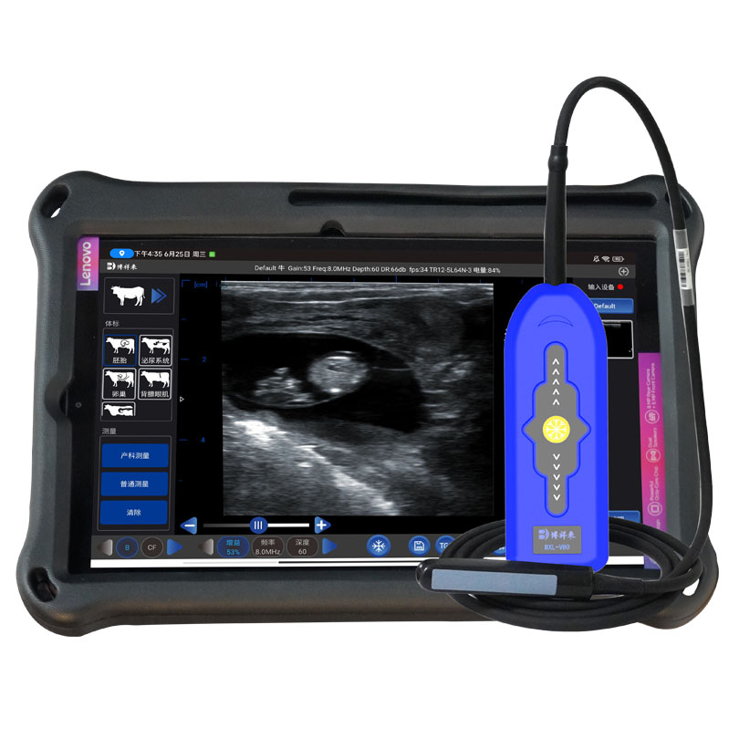
By examining vascular activity around the corpus luteum (CL), veterinarians can obtain additional information when evaluating reproductive status and early pregnancy.
So, how does colour Doppler pregnancy diagnosis in cows work? What role does corpus luteum blood flow play? And how can modern veterinary ultrasound equipment improve cattle reproductive examinations?
This article explores the technology, its practical applications, and the advantages of professional BOXERLY veterinary ultrasound systems.
1. What Is Colour Doppler Ultrasound in Cattle Pregnancy Diagnosis?
Colour Doppler ultrasound is an advanced imaging technology that detects and displays blood flow within tissues and blood vessels.
Unlike conventional B-mode ultrasound, which mainly displays anatomical structures, colour Doppler provides additional information about vascular activity.
In bovine reproductive examinations, this technology is particularly useful for evaluating:
Corpus luteum blood flow.
Ovarian vascular activity.
Uterine blood circulation.
Changes in reproductive tissues during early pregnancy.
Blood-flow patterns associated with different reproductive conditions.
Why is this important?
The corpus luteum is an important ovarian structure that produces progesterone, a hormone essential for maintaining pregnancy in cattle.
Following ovulation, the corpus luteum develops and becomes an important source of progesterone. Its structure and vascular activity change during the reproductive cycle.
During early pregnancy, the corpus luteum is generally maintained to support pregnancy. If pregnancy does not occur, the reproductive system undergoes changes that eventually lead to luteal regression.
Colour Doppler ultrasound allows veterinarians to examine blood-flow signals within the corpus luteum, providing additional information for reproductive assessment.
2. Why Is Corpus Luteum Blood Flow Important for Early Pregnancy Diagnosis in Cows?
The corpus luteum (CL) plays a central role in cattle reproductive physiology.
After ovulation, the corpus luteum develops from the ovulated follicle. Its function is to produce progesterone, which supports the uterine environment required for embryo development and pregnancy maintenance.
Blood supply is essential for normal corpus luteum development and function.
Research and practical veterinary applications have demonstrated that luteal vascularity can provide useful information when evaluating ovarian function and early pregnancy status.
With colour Doppler imaging, veterinarians can examine the distribution and intensity of blood-flow signals within the corpus luteum.
<Insert Image: Colour Doppler ultrasound image showing corpus luteum blood flow in a cow>
Potential benefits of examining corpus luteum blood flow include:
1. Evaluating luteal vascularity
Colour Doppler helps visualize blood-flow signals within the corpus luteum, supporting the assessment of its vascular development.
2. Supporting early pregnancy assessment
Changes in luteal blood flow may provide additional information when evaluating cows after artificial insemination.
3. Improving reproductive monitoring
Repeated ultrasound examinations allow veterinarians to monitor ovarian structures and reproductive changes over time.
4. Supporting more informed clinical decisions
Combining B-mode anatomical imaging with Doppler blood-flow information provides a more comprehensive examination than relying on anatomical images alone.
It is important to understand that corpus luteum blood flow is an additional diagnostic indicator, not independent proof of pregnancy. A reliable diagnosis should consider the complete reproductive examination, including uterine findings and embryonic development when visible.
3. Can Colour Doppler Ultrasound Detect Pregnancy in Cows at 21 Days?
One of the most interesting applications of advanced bovine ultrasound technology is early pregnancy assessment around 21 days after artificial insemination.
For cattle farmers, earlier pregnancy information can help improve breeding schedules and reduce unnecessary waiting periods.