Bovine Luteal Perfusion Pregnancy Diagnosis: Using Color Doppler Ultrasound for Early Pregnancy Detection in Cattle

Bovine Luteal Perfusion Pregnancy Diagnosis: Using Color Doppler Ultrasound for Early Pregnancy Detection in Cattle
1. What Is Bovine Luteal Perfusion Pregnancy Diagnosis?
Early pregnancy diagnosis is an essential part of modern cattle reproductive management. For veterinarians and cattle reproduction specialists, identifying pregnancy as early as possible can help improve breeding efficiency and reduce unnecessary reproductive costs.
One advanced approach is bovine luteal perfusion pregnancy diagnosis, which uses Color Doppler ultrasonography to evaluate blood circulation within the corpus luteum (CL) of the ovary.
The corpus luteum is a temporary endocrine structure that develops after ovulation. It produces progesterone, a hormone essential for maintaining pregnancy in cattle.
When a cow becomes pregnant, embryonic signaling helps maintain the corpus luteum and its progesterone production. In non-pregnant cows, luteal regression normally occurs as part of the reproductive cycle.
These physiological differences can be reflected in the blood-flow characteristics of the corpus luteum.
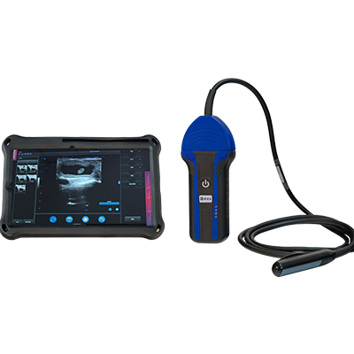
Using a veterinary ultrasound scanner equipped with Color Doppler technology, veterinarians can visualize luteal vascularization and evaluate blood perfusion patterns.
This technique provides valuable information for early pregnancy assessment, particularly around 20–22 days after artificial insemination (AI), depending on the examination protocol and animal population.
For professional cattle reproductive management, luteal blood-flow assessment represents an important application of modern veterinary Doppler ultrasound.
2. What Is the Corpus Luteum and Why Is Its Blood Flow Important?
The corpus luteum is formed from the ovarian follicle after ovulation.
Its primary function is to produce progesterone, which supports the uterine environment required for pregnancy establishment and maintenance.
The corpus luteum is a highly vascularized structure because hormone production requires an adequate blood supply.
In cattle, luteal development and regression are closely associated with changes in ovarian blood circulation.
Corpus Luteum in Pregnant Cows
Following successful fertilization, the developing embryo produces interferon tau (IFN-τ), which plays a key role in maternal recognition of pregnancy.
This signaling prevents the normal luteolytic process that would otherwise lead to corpus luteum regression.
Consequently, the corpus luteum is maintained, continuing to produce progesterone.
Color Doppler ultrasound may reveal relatively greater luteal blood perfusion in pregnant cows compared with non-pregnant animals during the early post-insemination period.
Corpus Luteum in Non-Pregnant Cows
When pregnancy does not occur, the uterine luteolytic mechanism generally causes the corpus luteum to regress.
During this process, luteal structure, hormone production, and blood-flow characteristics change.
Reduced luteal vascularization may therefore provide useful information when assessing whether a cow has established pregnancy.
However, luteal blood flow is an indirect indicator. It must be interpreted alongside reproductive history, ovarian findings, and appropriate follow-up examinations.
3. How Does Color Doppler Ultrasound Evaluate Luteal Blood Perfusion?
Color Doppler ultrasound is an imaging technology that displays blood-flow information in real time.
Unlike conventional B-mode ultrasound, which primarily shows anatomical structures in grayscale, Color Doppler adds color-coded information representing blood movement within tissues.
For bovine luteal perfusion assessment, the examination generally involves the following steps.
Step 1: Identify the Corpus Luteum
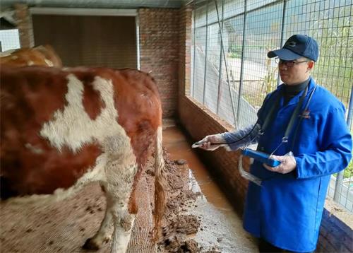
The veterinarian performs a transrectal ultrasound examination using a professional rectal linear probe.
In B-mode imaging, the ovaries are examined to identify the corpus luteum.
The examiner evaluates its location, size, shape, and internal appearance.
Step 2: Activate Color Doppler Mode
After identifying the corpus luteum, the veterinarian activates Color Doppler imaging.
Blood vessels and vascular signals within the luteal tissue become visible as color-coded areas on the ultrasound screen.
The distribution and extent of these signals provide information about luteal vascularization.
Step 3: Evaluate Blood-Flow Distribution
The examiner observes the blood-flow signals in different regions of the corpus luteum.
Important observations may include:
Presence of detectable blood-flow signals.
Distribution of peripheral vascularization.
Distribution of central vascularization.
Relative extent of perfused tissue.
Changes in luteal blood-flow patterns.
Some research protocols use image-analysis software to calculate the percentage of luteal tissue area containing Doppler blood-flow signals.